Nikos Priovoulos1, Icaro Agenor Ferreira de Oliveira1, Benedikt Poser2, David G Norris3,4, and Wietske van der Zwaag1
1Spinoza Center, Amsterdam, Netherlands, 2Maastricht University, Maastricht, Netherlands, 3Donders Institute for Brain, Cognition and Behaviour, Radboud University Nijmegen, Nijmegen, Netherlands, 4Erwin L. Hahn Institute for MRI, University of Duisburg-Essen, Essen, Germany
1Spinoza Center, Amsterdam, Netherlands, 2Maastricht University, Maastricht, Netherlands, 3Donders Institute for Brain, Cognition and Behaviour, Radboud University Nijmegen, Nijmegen, Netherlands, 4Erwin L. Hahn Institute for MRI, University of Duisburg-Essen, Essen, Germany
Arterial-Blood-Contrast can be created at submillimiter-resolution at 7T by suppressing the grey matter signal using magnetization transfer. We found good localization of Arterial-Blood-Contrast compared to BOLD that may be promising for future high-resolution applications.
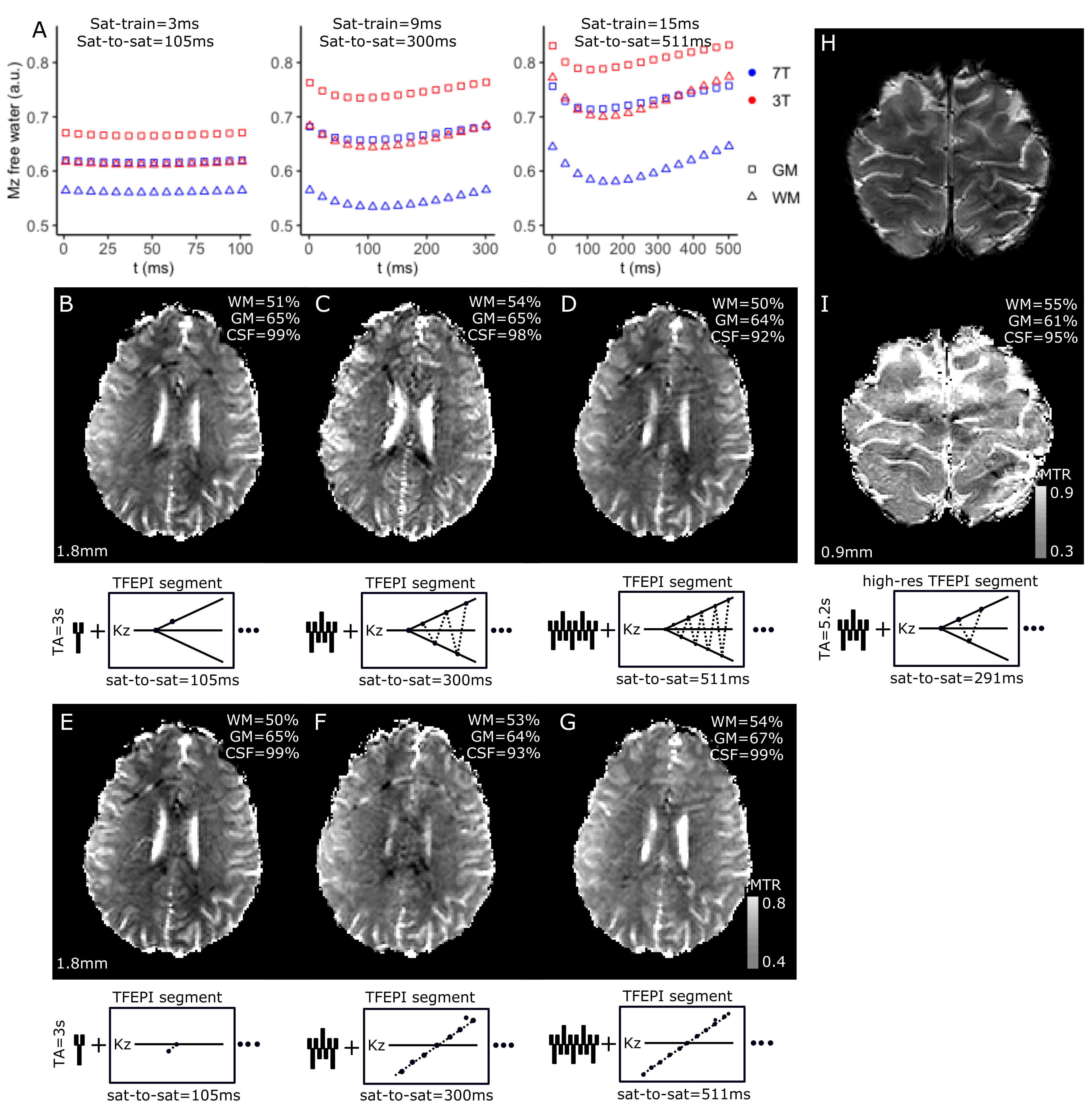
Figure 2: MT-weighting optimization for 7T. A, Simulation of Mz between saturations for the 3 schemes. Given the same RF, higher saturation can be achieved at 7T when longer delays between saturation trains are introduced. This makes high-resolution MT-weighted EPI feasible. B-G, in-vivo low-resolution TFEPI for center-slice-out (B-D) and linear (E-G) schemes. H-I, MT-weighted and MTR slice of the submillimeter TFEPI. The task data were collected with C and H.
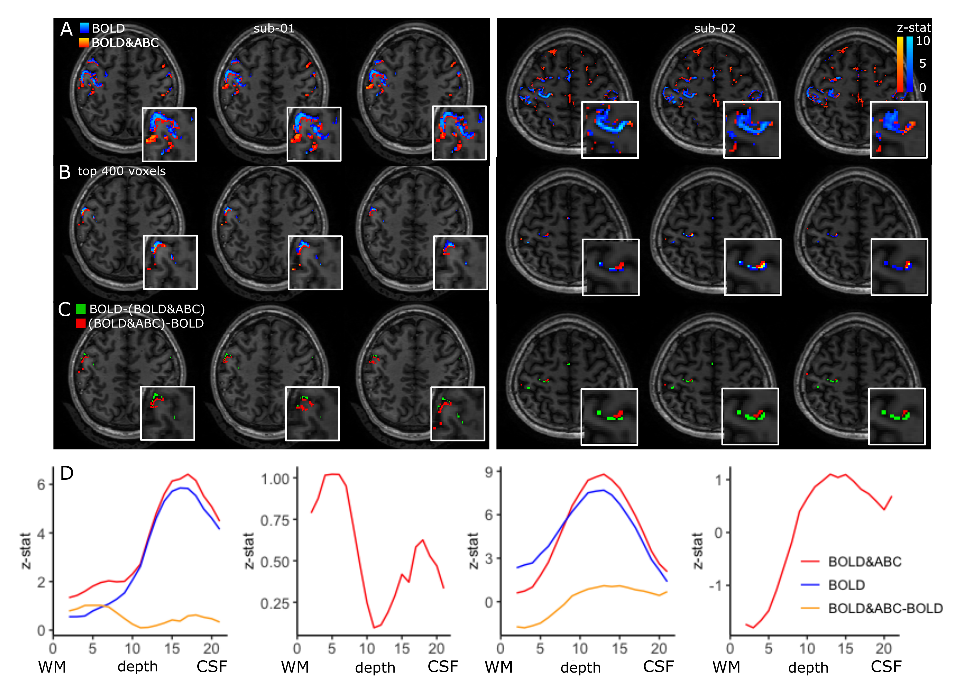
Figure 4: BOLD vs BOLD-and-ABC-weighted comparison (TFEPI 0.9mm isotropic). A, Successive slices of activation (blue/red-yellow: activation; green/red: difference between clusters) for two participants. B, 400 voxels with highest activation. C, Spatial difference between the BOLD and BOLD-and-ABC (top 400 voxels only). Note that adding ABC contrast seems to shift the activation deeper in the cortex compared to BOLD. D, Laminar profiles. Subtracting BOLD from BOLD-and-ABC shows increased cortical specificity and even a double-peak profile for one participant.