Hong-Hsi Lee1, Els Fieremans1, Susie Y Huang2, Qiyuan Tian2, and Dmitry S Novikov1
1New York University School of Medicine, New York, NY, United States, 2Department of Radiology, A. A. Martinos Center for Biomedical Imaging, Massachusetts General Hospital, Charlestown, MA, United States
1New York University School of Medicine, New York, NY, United States, 2Department of Radiology, A. A. Martinos Center for Biomedical Imaging, Massachusetts General Hospital, Charlestown, MA, United States
We perform numerical simulations and in vivo diffusion MRI measurements of strong gradients on a connectome scanner to demonstrate the localization regime of diffusion and estimate soma size in cortical brain gray matter of healthy subjects.
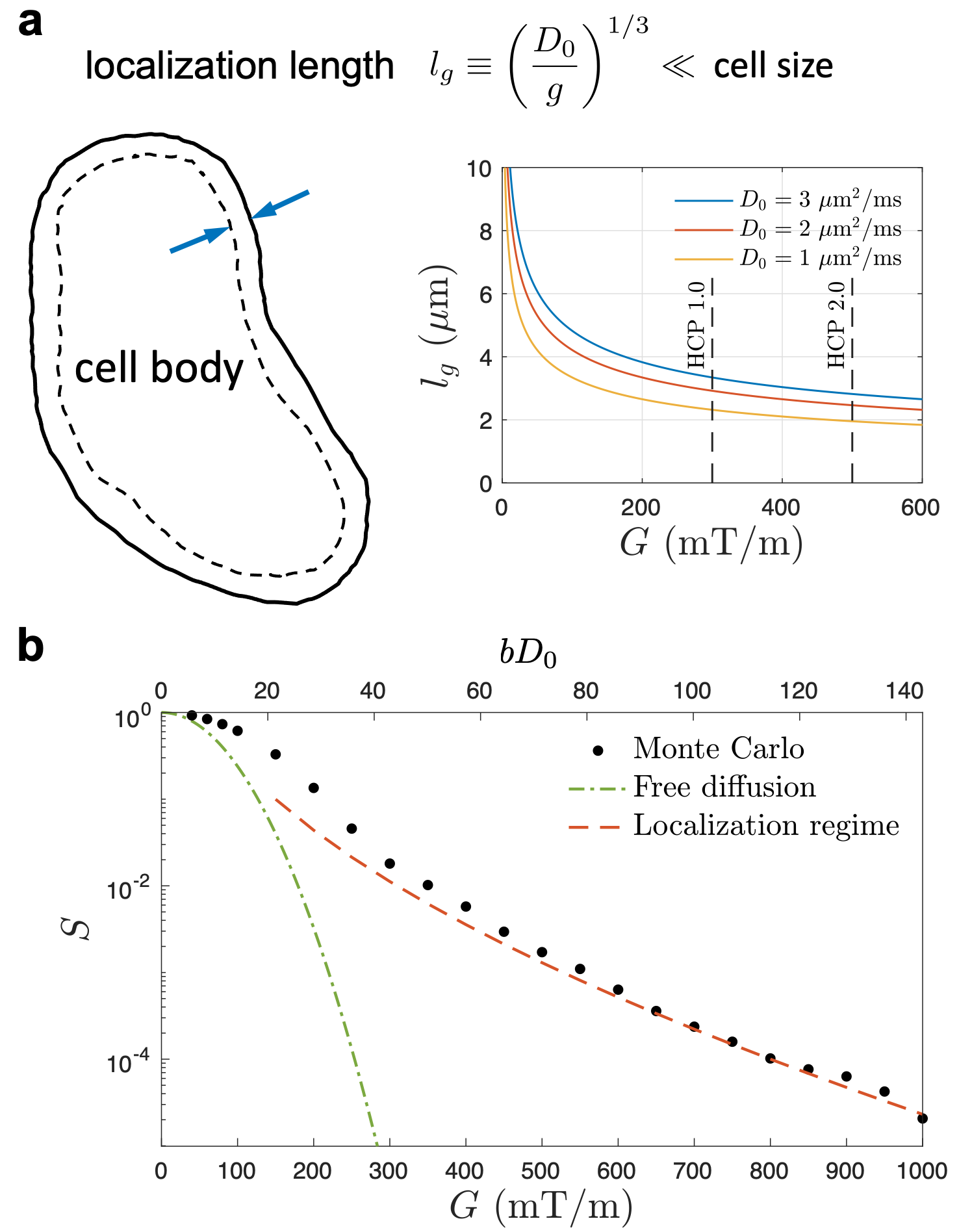
Fig. 1. a. By applying strong gradients G (Larmor gradient g=γG), magnetization far from the boundaries (cell membranes) vanishes, and that near the boundaries within a thickness of localization length lg contributes to measured signal. This so-called localization regime emerges when lg << diffusion length and cell dimension. b. Numerical simulation of diffusion inside an impermeable sphere (D0=3 μm2/ms) demonstrates the localization regime at strong gradients (dashed line). The signal decay in localization regime is much slower than the free diffusion (dotted-dashed line).
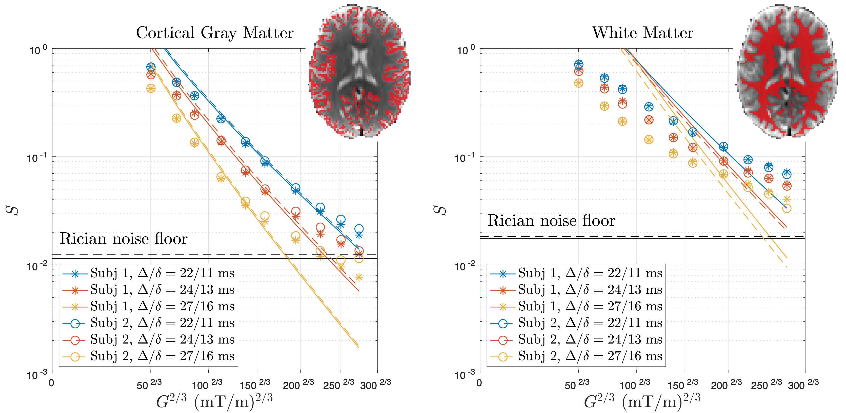
Fig. 3. dMRI measurements by using wide pulse sequence of strong gradients at 3 different diffusion times in 2 healthy subjects demonstrates the localization regime in the cortical brain gray matter, where diffusion signals coincide with the theory in Eq. (2) and yield a soma size estimation (Fig. 4). In contrast, diffusion signals in the brain white matter are not compatible to theory of localization regime since the axon size ~1 μm is smaller than the localization length lg = 3.4-6.0 μm in experiments.