Maëliss Jallais1, Pedro L. C. Rodrigues1, Alexandre Gramfort1, and Demian Wassermann1
1Université Paris-Saclay, Inria, CEA, Palaiseau, France
1Université Paris-Saclay, Inria, CEA, Palaiseau, France
Based on a new
forward model of brain grey matter, we extract summary statistics
from an acquired diffusion signal and estimate the tissue parameters
that best describe it, along with a full posterior distribution over
the parameter space, using a likelihood-free inference based
algorithm.
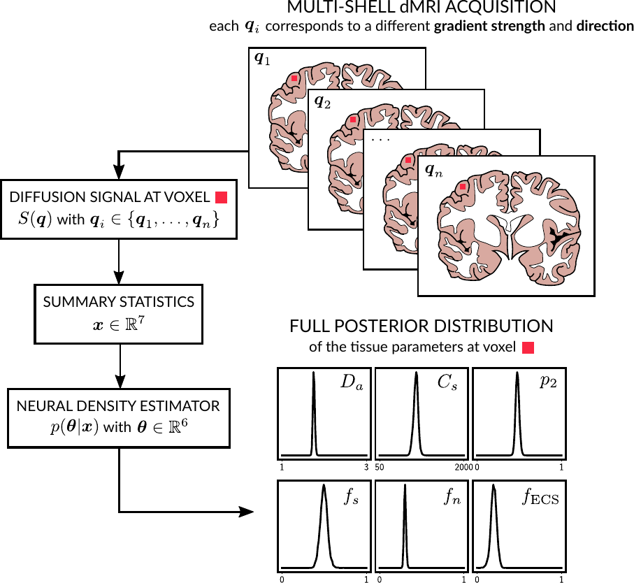
Visual abstract. On the top right we illustrate a multi-shell dMRI acquisition. Based on the proposed 3-compartment model, we then extract summary statistics from it. Applying a neural density estimator we can both estimate the tissue parameters and their full posterior distribution.
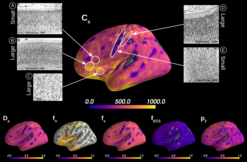
Microstructural measurements averaged over 31 HCP MGH subjects. We deemed stable measurements with a z-score larger than 2, where the standard deviation on the posterior estimates was estimated through our LFI fitting approach. In comparing with Nissl-stained cytoarchitectural studies we can qualitatively evaluate our parameter Cs: Broadmann area 44 (A) has smaller soma size in average than area 45 (B)13; large von Economo neurons predominate the superior anterior insula (C)12; precentral gyrus (E) shows very small somas while post-central (D) larger ones14.