Marco Palombo1, Daniel C. Alexander1, and Hui Zhang1
1Centre for Medical Image Computing, University College London, London, United Kingdom
1Centre for Medical Image Computing, University College London, London, United Kingdom
Statistics for a comprehensive set of morphological features derived from ~3,500 brain cells across species. Implications for biophysical modelling of gray matter discussed, enabling the design of more sensible models and suitable data acquisitions.
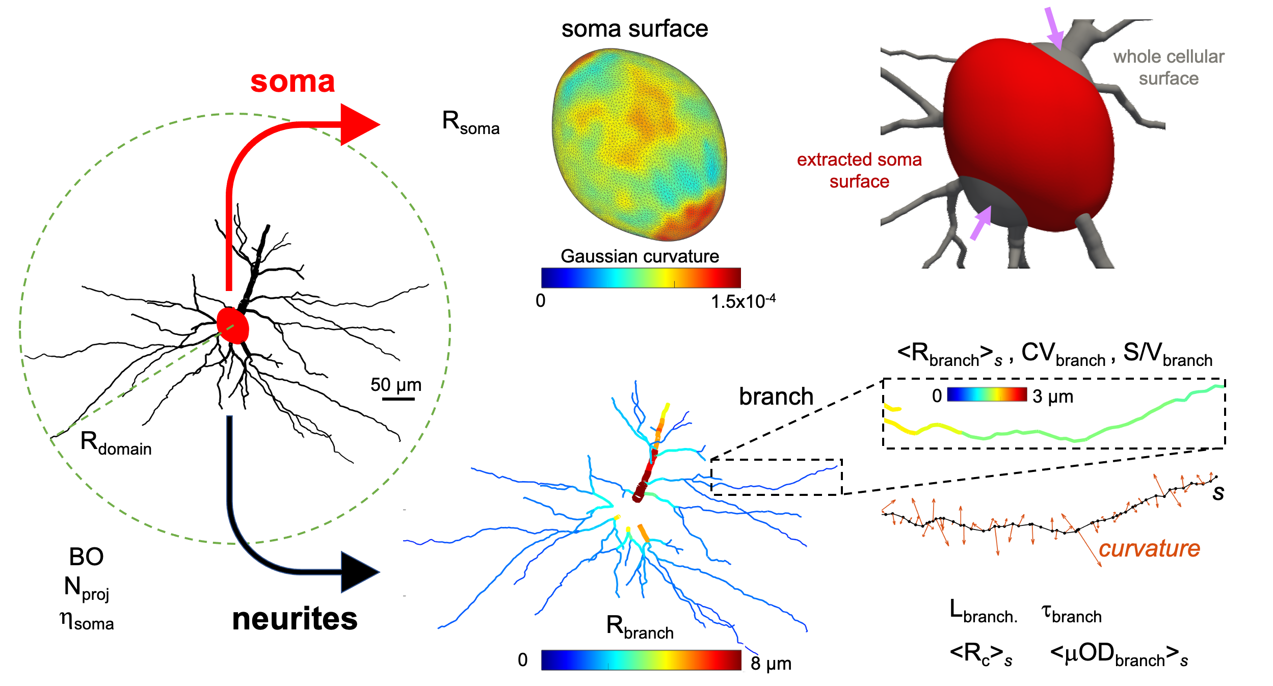
Fig.2 Illustration of the morphological features investigated for an exemplar cell. We estimated general features of the whole structure and separated soma from neurites, processing them individually to estimate a set of other relevant features (see Methods). Additionally, we display the Gaussian curvature of the soma surface to show that it is a non-spherical geometry (always positive but not constant). A limitation of the current approach (and the majority of existing tools23,24) is the slightly inaccurate definition of the soma surface, as shown in the top right corner (arrows).
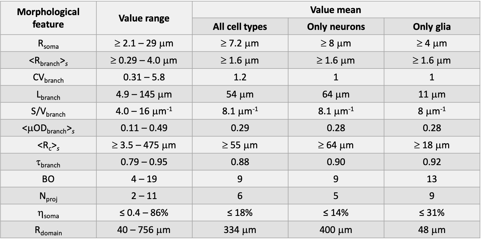
Tab.2 Reference values for all the morphological features of neuronal and glial cells. The ranges and mean values obtained from the whole dataset investigated (N = 3,448) are reported, together with mean values for only neurons and glia. The ‘≥’ and ‘≤’ are used when the estimated value of the corresponding feature may be slightly (on average <20%) under- or over-estimated, respectively, given the known limitations of the approach used (e.g., see Fig.2).