Mariam Andersson1,2, Marco Pizzolato1,2,3, Hans Martin Kjer1,2, Henrik Lundell1, and Tim B. Dyrby1,2
1Danish Research Centre for Magnetic Resonance, Hvidovre, Denmark, 2Technical University of Denmark, Kgs. Lyngby, Denmark, 3Signal Processing Laboratory (LTS5), École Polytechnique Fédérale de Lausanne, Lausanne, Switzerland
1Danish Research Centre for Magnetic Resonance, Hvidovre, Denmark, 2Technical University of Denmark, Kgs. Lyngby, Denmark, 3Signal Processing Laboratory (LTS5), École Polytechnique Fédérale de Lausanne, Lausanne, Switzerland
In axons from a complex crossing fiber region of the primate brain, powder averaging of the diffusion MRI signal removes orientation bias in axon diameter estimates. The wide range of diameters present on the subvoxel scale indicates a need for methods with broad sensitivity to diameter.
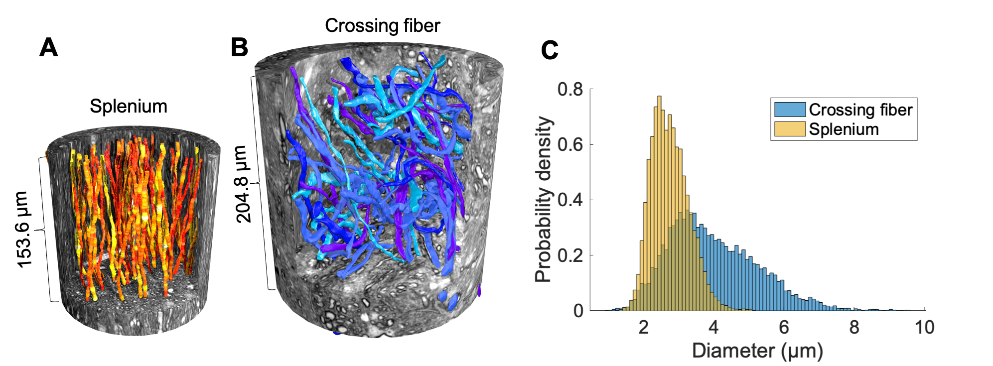
Figure 1. 3D reconstructions of A) 54 splenium axons (segmented at 75 nm resolution) and B) 58 crossing fiber axons (segmented at 500 nm resolution) in their respective XNH volumes. C) Combined 3D AD distributions over all measured diameters in the splenium (yellow) and crossing fiber region (blue).
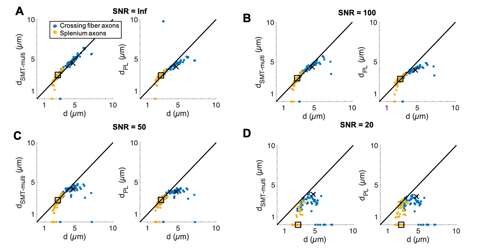
Figure 3. AD estimation in real axons. Estimated diameter from the SMT approach ($$$d_{SMT-multi})$$$ and PL approach ($$$D_{PL}$$$) vs the geometrical volume-weighted AD, $$$d$$$ for A) infinite SNR (ignoring the inherent MC noise), B) SNR = 100, C) SNR = 50 and D) SNR = 20, where fitting fails for the population of splenium axons. Square marker: volume-weighted AD of splenium axon population, cross marker: volume-weighted AD of crossing fiber population.