D Rangaprakash1 and Robert L Barry1
1Athinoula A. Martinos Center for Biomedical Imaging, Massachusetts General Hospital and Harvard Medical School, Charlestown, MA, United States
1Athinoula A. Martinos Center for Biomedical Imaging, Massachusetts General Hospital and Harvard Medical School, Charlestown, MA, United States
Functional MRI is confounded by the HRF, but the nature of this confound in the spinal cord is not well understood. We observed cervical cord HRF to have 6–9% within- and between-subjects variability in the gray matter and 3–5% in the white matter.
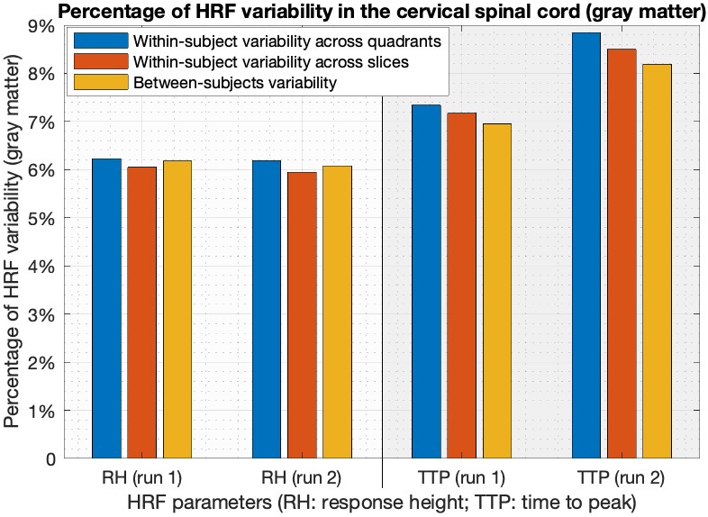
Figure-4. Percentage of HRF variability in the gray matter. In both the runs, RH consistently showed about 6% variability across quadrants, spinal levels (=slices) and subjects. TTP showed higher variability than RH (7–9%) but seemed less consistent across runs. This lesser consistency could perhaps be due to the poorer temporal resolution of our data (VAT=2.08s) (this is also a limitation of the study), as TTP estimates are quantified in units of VAT.
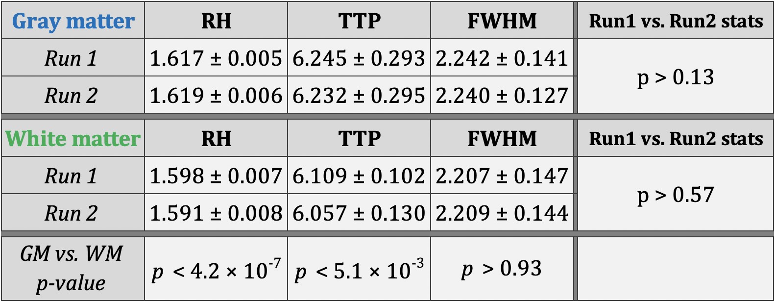
Figure-3. Simple statistics of response height (RH), time-to-peak (TTP) and full-width at half max (FWHM) in the cervical spinal cord. Mean ± standard deviation of HRF parameters aggregated across all quadrants, levels and subjects is shown for both runs. The last column comparing runs 1 and 2 shows that aggregated HRF parameters are consistent across runs. The last row comparing gray (GM) and white matter (WM) shows that RH and TTP are significantly higher in the GM.