Shiva Shahrampour1, Benjamin De Leener2, Mahdi Alizadeh1, Devon Middleton1, Laura Krisa1, Adam Flanders1, Scott Faro1, Julien Cohen-Adad2, and Feroze Mohamed1
1Thomas Jefferson University, Philadelphia, PA, United States, 2Polytechnique Montreal, Montreal, QC, Canada
1Thomas Jefferson University, Philadelphia, PA, United States, 2Polytechnique Montreal, Montreal, QC, Canada
We were able to automatically delineate various white matter tracts of the healthy pediatric spinal cord. The normative DTI indices associated with those tracts were quantified. The effect of age on the maturation of the tracts was studied and showed significant difference using MD, RD and AD.
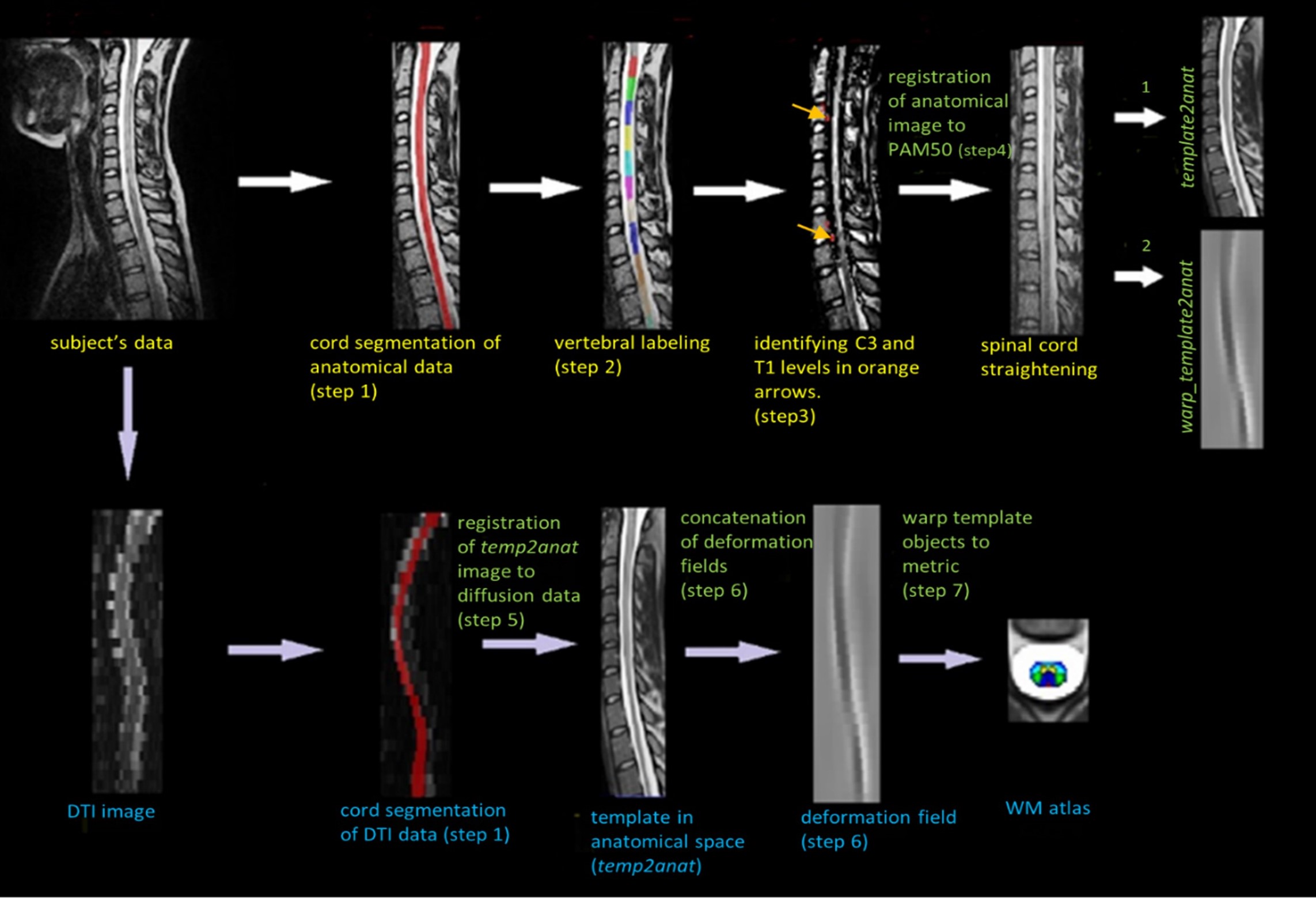
Figure 1. Overview of template registration
pipeline. Initially, T2-weighted scans are registered to the template (top
row). DTI data acquired during the same scan session are then registered to the
anatomical data and PAM 50 objects are warped to diffusion data (bottom row) to
generate the pediatric WM spine atlas.
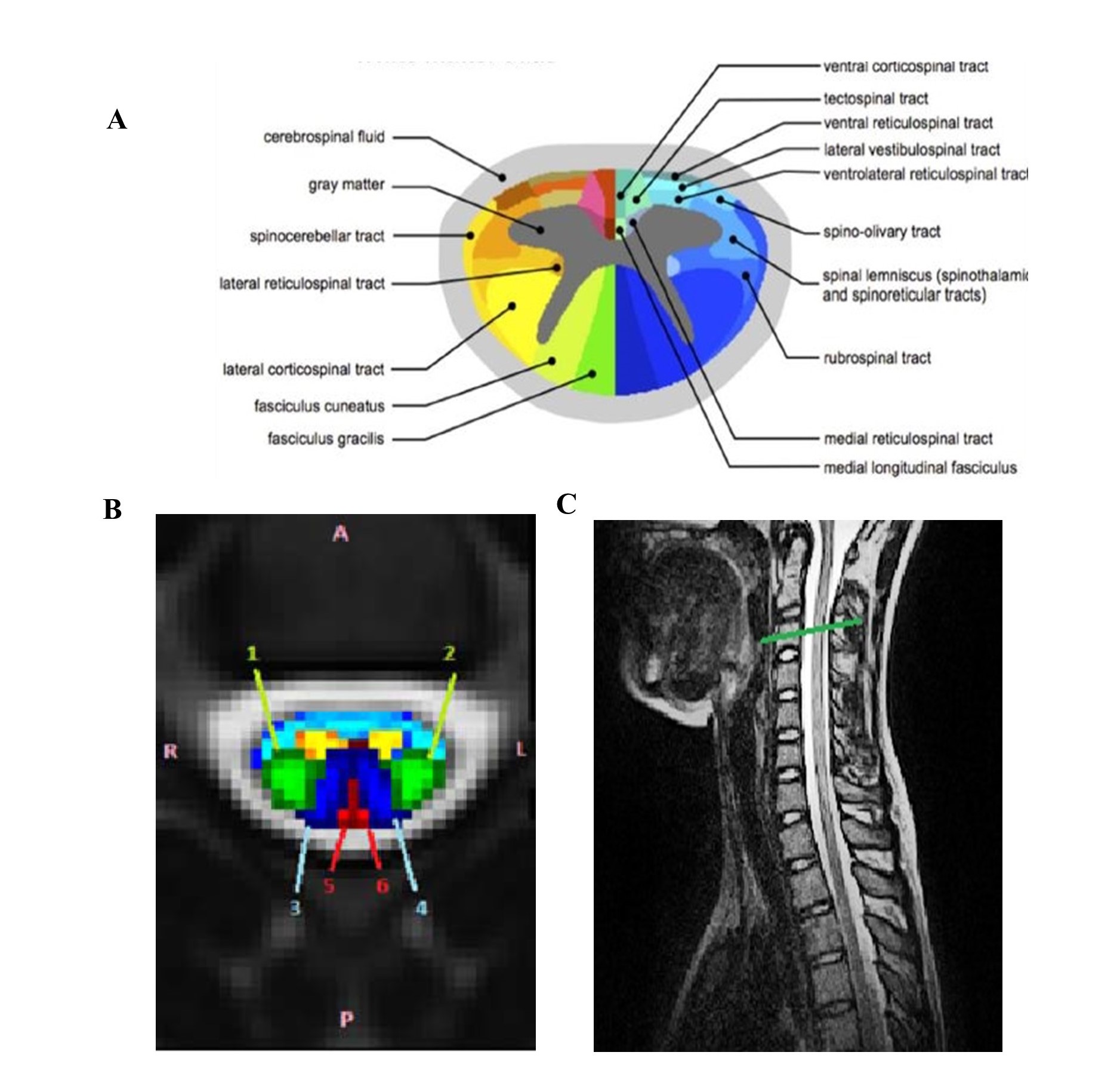
Figure 3.
Spinal cord WM atlas: A. An
atlas of spinal white matter tracts derived from the Gray’s Anatomy atlas B. Generated white matter atlas of
pediatric spinal cord overlaid on a b0 image. The selected tracts are labeled
with multiple colors. C. The C3 level
is marked in green color on the sagittal T2-weighted scan.