Arvin Arani1, Christopher G. Schwarz1, Matthew C. Murphy1, Joshua D. Trzasko1, Jeffrey L. Gunter1, Matthew L. Senjem1, Heather J. Wiste1, Kiaran P. McGee1, Matthew A. Bernstein1, John Huston III1, and Clifford R. Jack Jr.1
1Mayo Clinic, Rochester, MN, United States
1Mayo Clinic, Rochester, MN, United States
This
study shows that left-right intensity asymmetries are system specific,
systematic, can mimic disease and create diagnostic uncertainty, and that they
impact multiple sequences (T1-weighted and FLAIR).
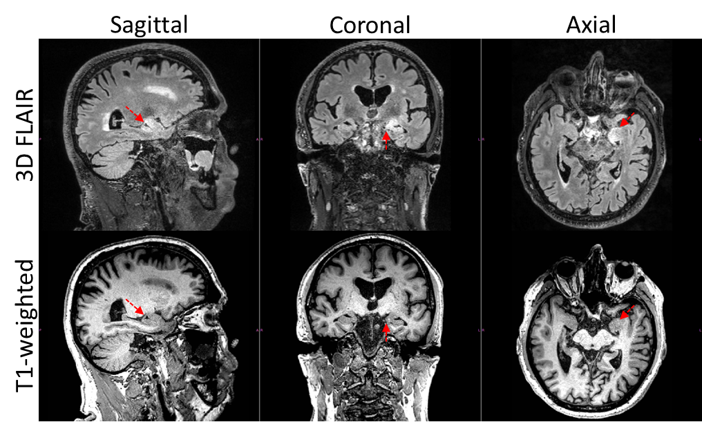
Localized regions of
inhomogeneity observed on the same patient in both 3D FLAIR and T1-w diagnostic
images. In this case the localized region (red arrows) was thought to be
suspicious of potential herpes encephalitis and follow-up imaging was
conducted.
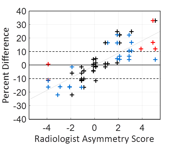
Two experienced
radiologist’s asymmetry scores (assessment localized to the hippocampus), are
plotted against the percent difference in intensity measured in the hippocampus
(left side > right side) with automated atlas-based segmentation. The same
30 image volumes from 30 different patients were scored. The color of the data
points corresponds to the response of each radiologist to the question: “Is
clinical follow-up required?” where: definitely not (black +),
uncertain (blue +), definitely yes (red +).