Jun Chen1, Junjie Ma1, Crystal E Harrison1, James Ratnakar1, Zungho Zun2, Jeff Liticker1, Galen D Reed3, Avneesh Chhabra4, Thomas Jue5, Craig R Malloy1,3,6, and Jae Mo Park1,4,7
1AIRC, UT Southwestern Medical Center, Dallas, TX, United States, 2The Developing Brain Institute, Children’s National Hospital, Washington, DC, United States, 3GE, Chicago, IL, United States, 4Radiology, UT Southwestern Medical Center, Dallas, TX, United States, 5Biochemistry and Molecular Medicine, UC Davis, Davis, CA, United States, 6Internal Medicine, UT Southwestern Medical Center, Dallas, TX, United States, 7Electrical and Computer Engineering, University of Texas at Dallas, Richardson, TX, United States
1AIRC, UT Southwestern Medical Center, Dallas, TX, United States, 2The Developing Brain Institute, Children’s National Hospital, Washington, DC, United States, 3GE, Chicago, IL, United States, 4Radiology, UT Southwestern Medical Center, Dallas, TX, United States, 5Biochemistry and Molecular Medicine, UC Davis, Davis, CA, United States, 6Internal Medicine, UT Southwestern Medical Center, Dallas, TX, United States, 7Electrical and Computer Engineering, University of Texas at Dallas, Richardson, TX, United States
This study demonstrates the feasibility of imaging skeletal muscle metabolism using hyperpolarized [1-13C]pyruvate and the sensitivity of in-vivopyruvate metabolism to exercise states
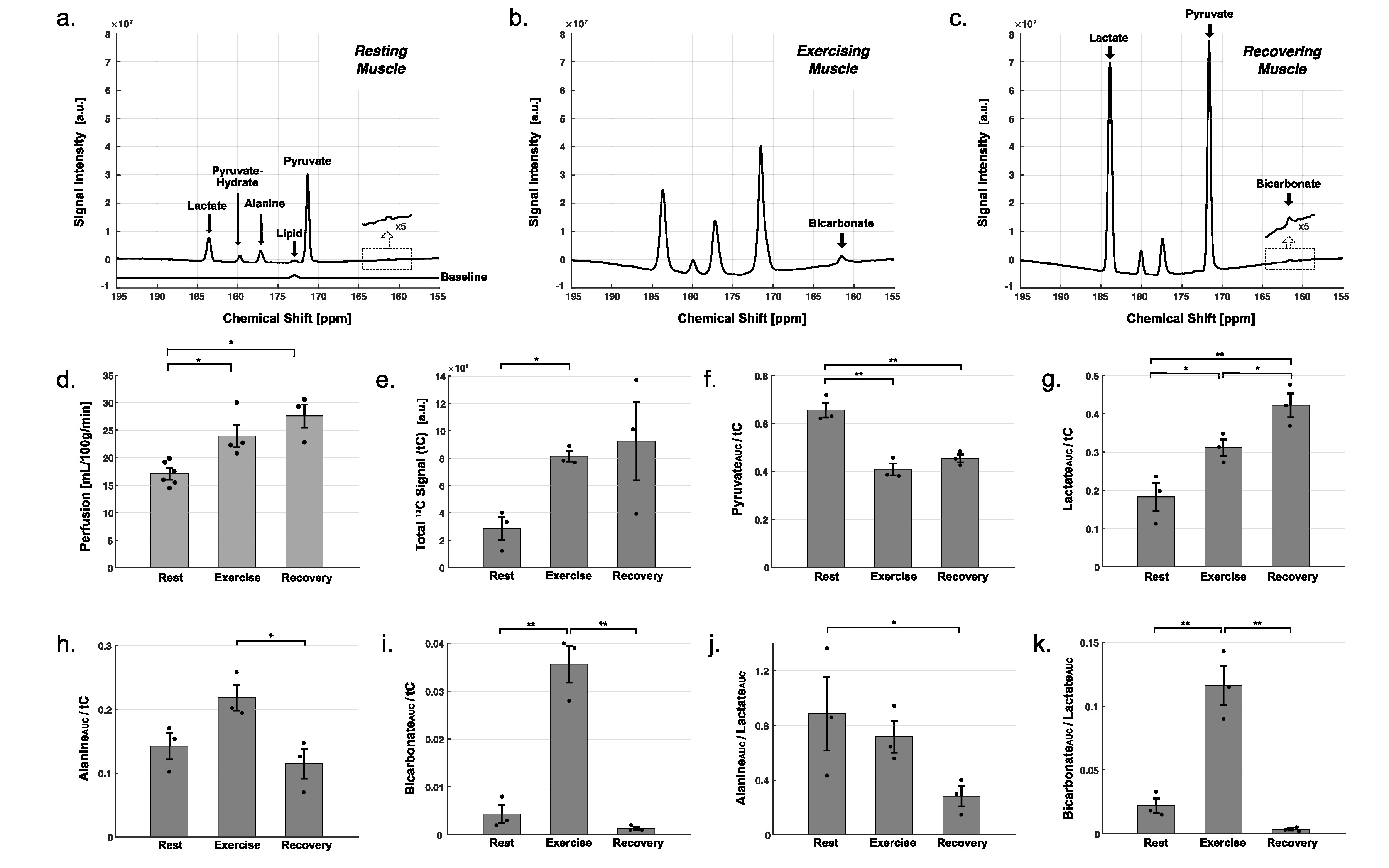
Effect of exercise in HP 13C MRS. Time-averaged 13C spectra over the first 90 seconds after bolus injection of HP [1-13C]pyruvate from a representative participant (a) at rest, (b) during exercise, and (c) recovery. (d) Spatially-averaged perfusion measured by 1H ASL from all the participants. (e) Total 13C signals (tC). Area under the curves (AUCs) for (f) [1-13C]pyruvate, (g) [1-13C]lactate, (h) [1-13C]alanine, and (i) [13C]bicarbonate, normalized by the tC AUC. AUCs of (j) alanine and (k) bicarbonate, normalized by the lactate AUC. (*: p<0.05, **: p<0.01).
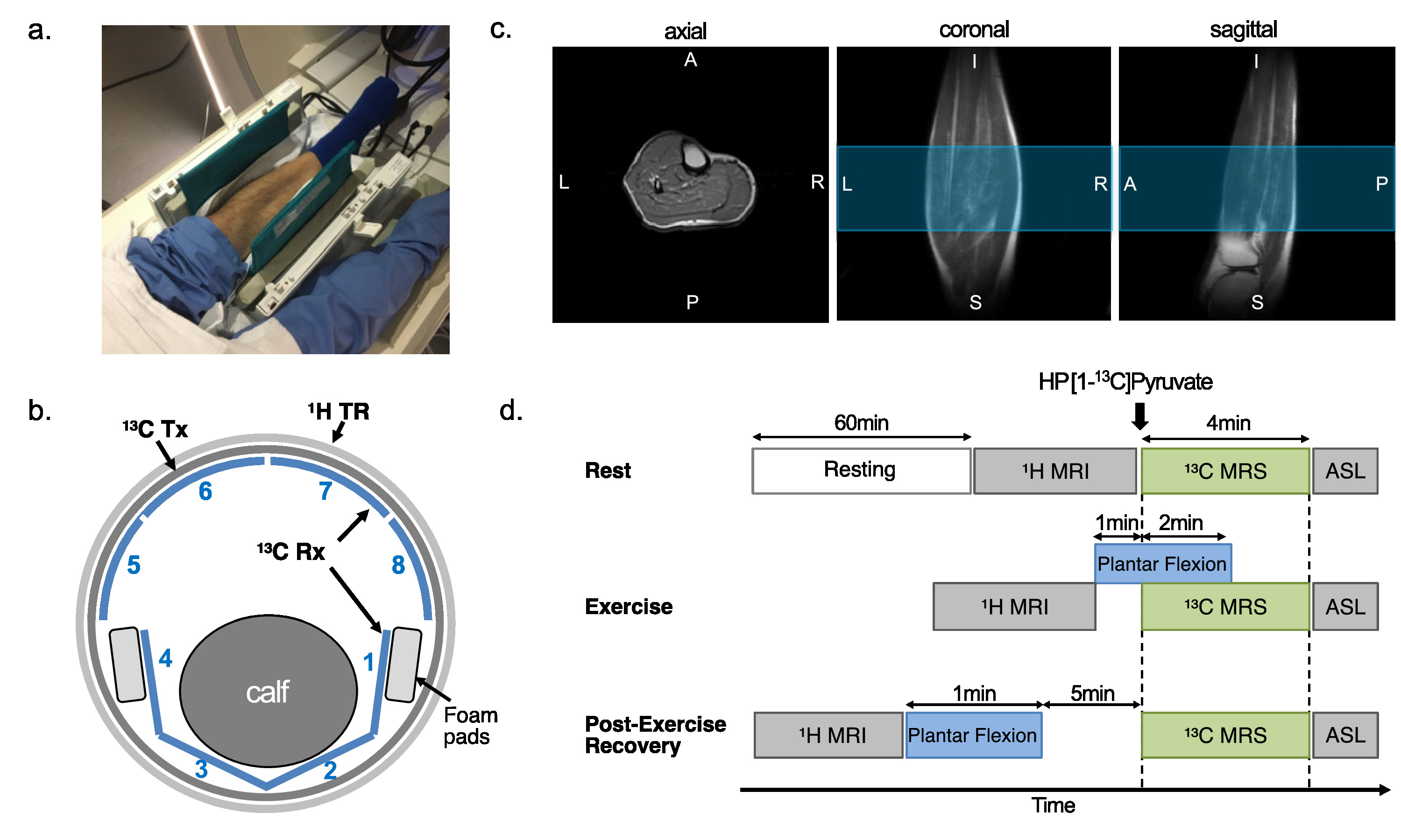
Experimental setup and study protocol. (a) Positioning calf muscle in a 13C/1H dual-frequency RF coil prior to connect anterior part of the coil. (b) Calf muscle was wrapped by the flexible posterior 13C receive arrays (channel #1-4). (c) Localized single-shot fast spin echo 1H images acquired using the RF coil. Blue region indicates the prescribed axial slab for 13C MRS (10-cm thick). (d) Dynamic 13C MRS was acquired at three metabolic states: rest, exercise, and recovery.