Helen Marshall1, Grace T Mussell1, Laurie J Smith1, Alberto M Biancardi1, Paul JC Hughes1, Andrew J Swift1, Smitha Rajaram1, Alison M Condliffe1, Guilhem J Collier1, Chris S Johns1, Nick D Weatherley1, Ian Sabroe2, and Jim M Wild1
1University of Sheffield, Sheffield, United Kingdom, 2Sheffield Teaching Hospitals, Sheffield, United Kingdom
1University of Sheffield, Sheffield, United Kingdom, 2Sheffield Teaching Hospitals, Sheffield, United Kingdom
129Xe ventilation MRI can provide additional
unique and valuable information in the evaluation of clinical presentations of
asthma, when undertaken as part of an MDT evaluation of severe disease.
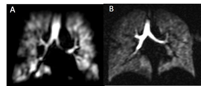
Figure
2: (A) shows
small to moderate sized ventilation defects in a symptomatic patient with
consistently normal spirometry. (B) shows relatively homogeneous
ventilation in a highly symptomatic patient.
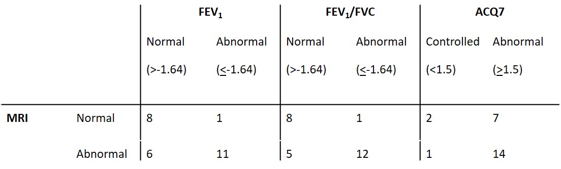
Table
3: Summary of subjects’ MRI classification, spirometry and ACQ7 scores. MRI
classified based on radiologists’ reports (normal = no or minor ventilation
defects, abnormal = substantial ventilation defects). Note:
ACQ7 score was available for 24 patients within 3 months of the scan.