Elianna Ada Bier1, Fawaz Alenezi2, Junlan Lu3, Joseph G Mammarappallil4, Bastiaan Driehuys4, and Sudarshan Rajagopal2
1Biomedical Engineering, Duke University, Durham, NC, United States, 2Division of Cardiology, Department of Medicine, Duke Univeristy, Durham, NC, United States, 3Medical Physics Graduate Program, Duke University, Durham, NC, United States, 4Radiology, Duke University, Durham, NC, United States
1Biomedical Engineering, Duke University, Durham, NC, United States, 2Division of Cardiology, Department of Medicine, Duke Univeristy, Durham, NC, United States, 3Medical Physics Graduate Program, Duke University, Durham, NC, United States, 4Radiology, Duke University, Durham, NC, United States
129Xe MRI/MRS can be used to non-invasively
detect pulmonary hypertension (PH). Here we extend these techniques by
evaluating imaging of 129Xe signal oscillations in red blood cells
in patients who have undergone right heart catheterization to determine PH
status.
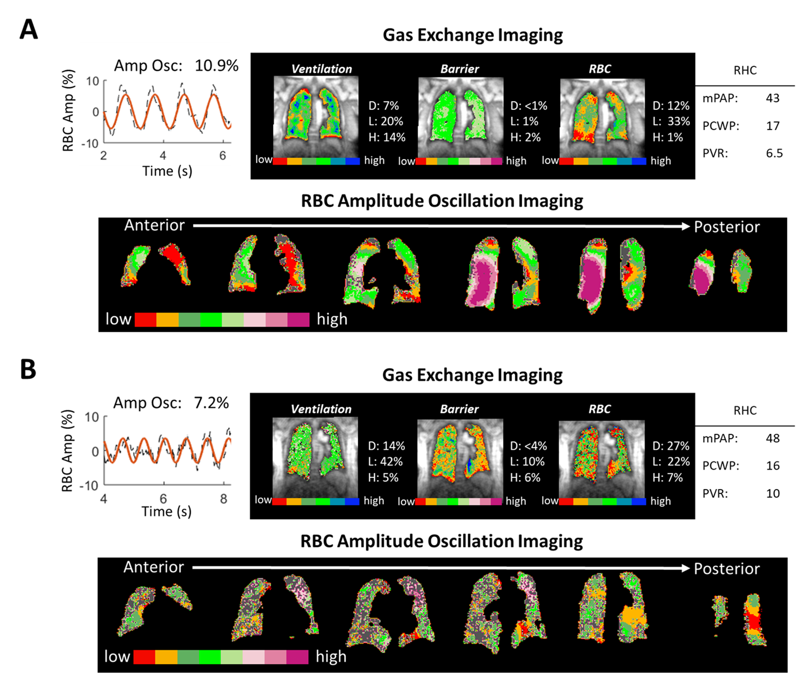
Figure 5. 129Xe MRI/MRS and RBC oscillation imaging
for 2 subjects with CpcPH and no extensive obstructive or interstitial disease.
The conventional algorithm classifies Subject A as no PH, and subject B as precapillary
PH. Oscillation imaging in both subjects indicates regions of both low and high
oscillations. Thus, imaging appears to correctly indicate the presence of both
pre- and postcapillary PH.
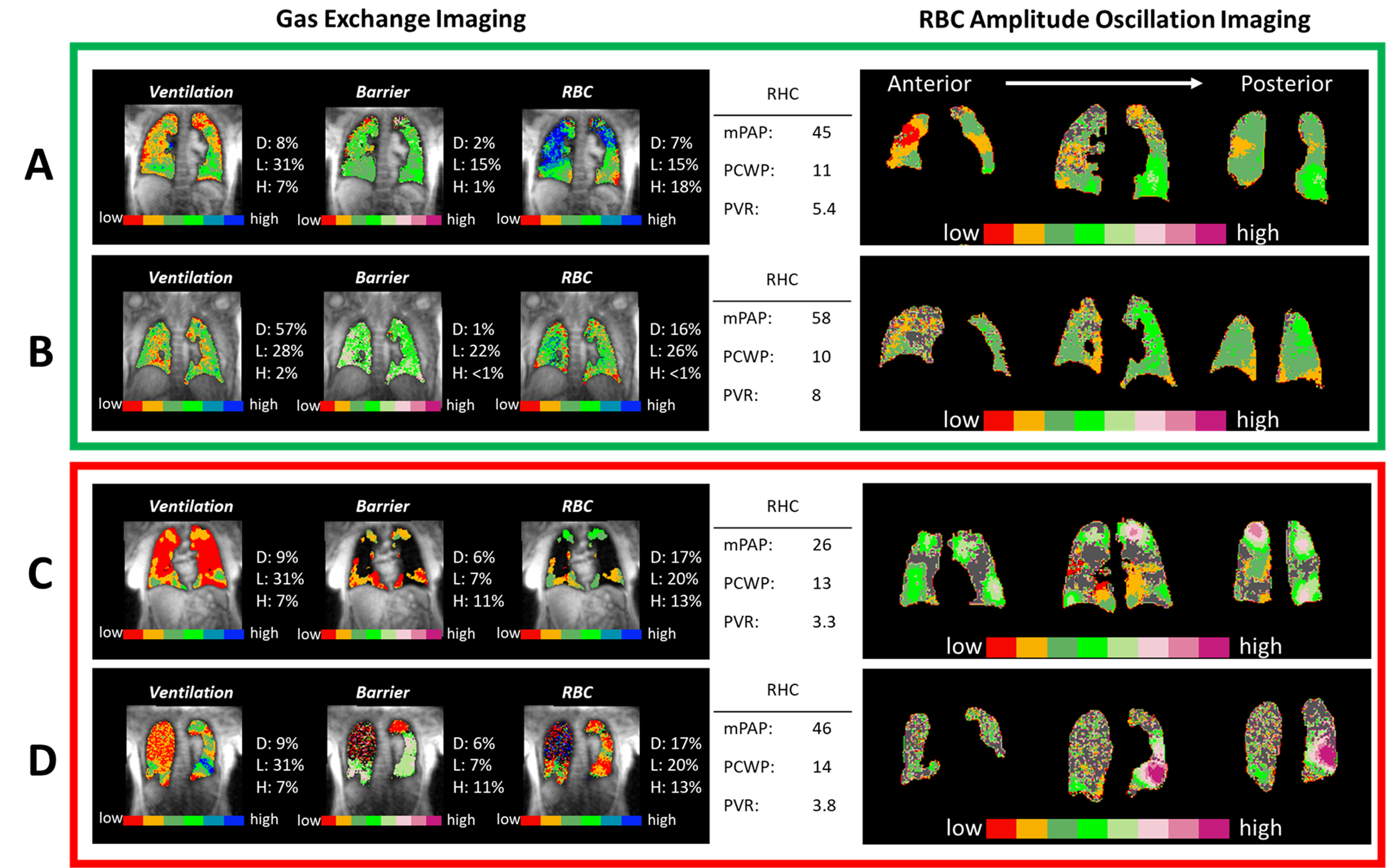
Figure 4. 129Xe gas exchange and RBC oscillation
imaging for test subjects classified by RHC as pre-capillary PH. Subjects A and
B exhibit RBC amplitude oscillations of 5.6%, and 5.4%, and are both properly
classified as PH-pre. These subjects also exhibit regions of low RBC
oscillations on imaging. Subject C and D have RBC oscillation amplitudes of 10.9%
and 9.3% leading the basic algorithm to incorrectly classify both classified as
no PH. These subjects have extensive parenchymal disease and oscillation
imaging is heterogeneous, suggesting PH may be in proportion to lung disease.