Agilo Luitger Kern1,2, Marcel Gutberlet1,2, Frank Wacker1,2, Jens Hohlfeld2,3,4, and Jens Vogel-Claussen1,2
1Institute of Diagnostic and Interventional Radiology, Hannover Medical School, Hannover, Germany, 2Biomedical Research in Endstage and Obstructive Lung Disease Hannover (BREATH), German Center for Lung Research (DZL), Hannover, Germany, 3Department of Clinical Airway Research, Fraunhofer Institute for Toxicology and Experimental Medicine, Hannover, Germany, 4Department of Respiratory Medicine, Hannover Medical School, Hannover, Germany
1Institute of Diagnostic and Interventional Radiology, Hannover Medical School, Hannover, Germany, 2Biomedical Research in Endstage and Obstructive Lung Disease Hannover (BREATH), German Center for Lung Research (DZL), Hannover, Germany, 3Department of Clinical Airway Research, Fraunhofer Institute for Toxicology and Experimental Medicine, Hannover, Germany, 4Department of Respiratory Medicine, Hannover Medical School, Hannover, Germany
A hyperpolarized 129Xe
stimulated echo pulse sequence was implemented. A study in healthy volunteers
and chronic obstructive pulmonary disease (COPD) patients showed a reduction of
stimulated echo signal in COPD, demonstrating diagnostic potential.
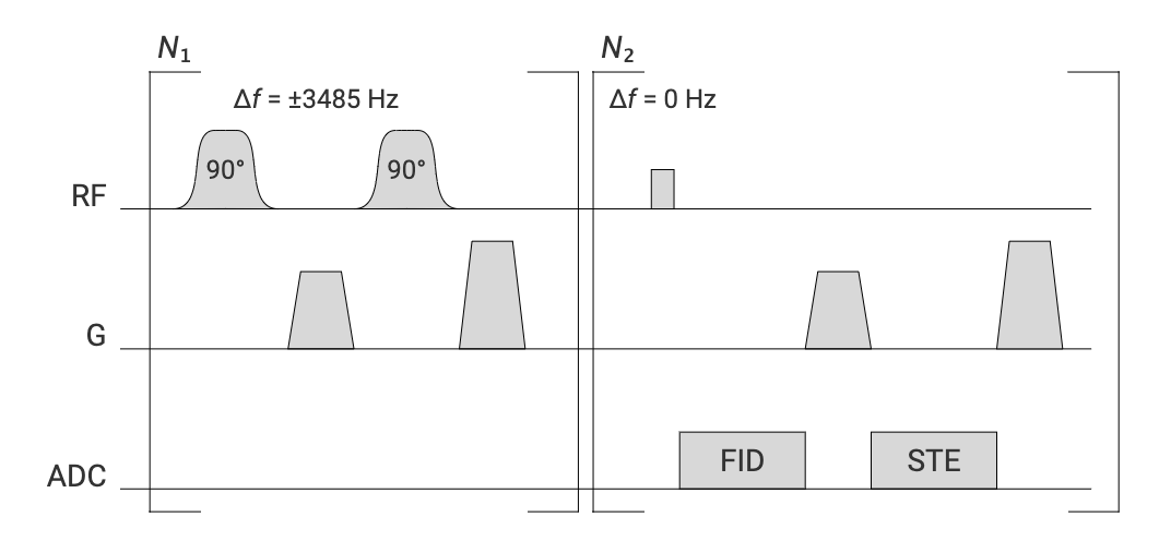
Figure 1. Pulse
sequence diagram. The phase difference of the two 90° pulses was chosen to be 0
at the given off-resonance frequency Δf. Fermi-shaped pulses are used
for selectively creating spatially modulated magnetization in the 129Xe
dissolved phase while providing a sufficient amplitude integral to achieve 90°
flip angles within the dissolved phase T2*. The whole sequence is
repeated as a control acquisition with inverted off-resonance frequency and
STEAM de- and rephasing gradients rotated by 90°.
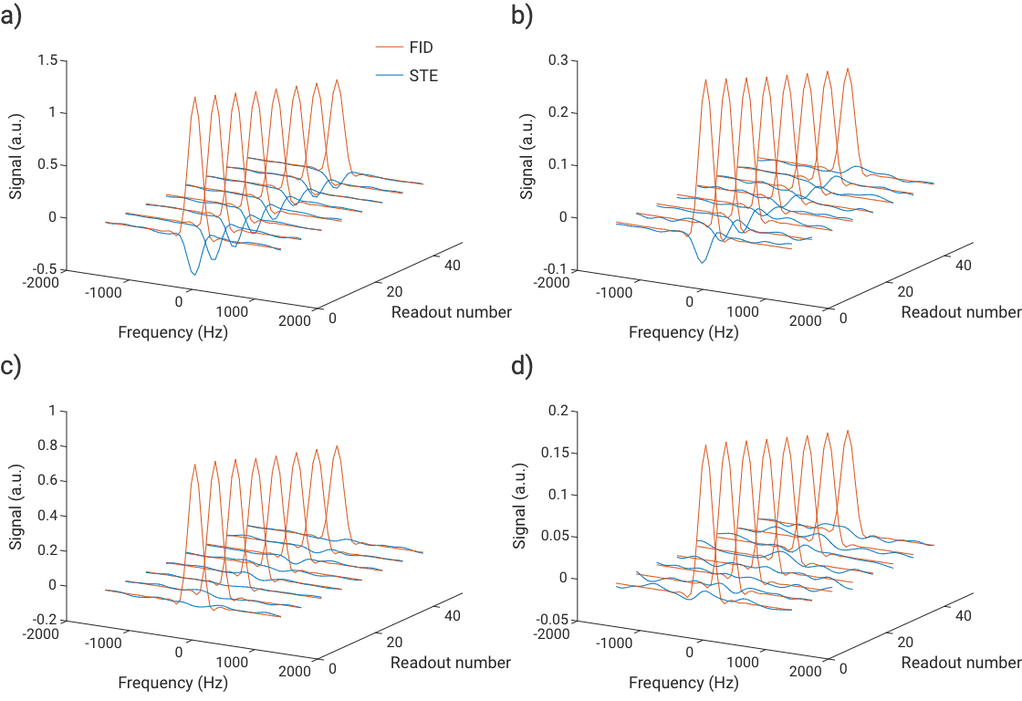
Figure 2.
Exemplary line-broadened spectra after off-resonance correction and phasing for
the FID signal from a) a healthy volunteer and b) a COPD patient after
excitation at +3485 Hz. Real part of FID signal shown in orange, STE signal in
blue and rescaled by factor 100. In a) as expected for pure TP excitation, an
approximate phase difference of π is found. In b) at later times a noticeable phase
change is observed, possibly due to signal contamination or motion. Control
acquisitions are shown for the HV in c) and the COPD patient in
d). Only small signals are observed
in the STE pathway.