Brice J Albert1, Peter J Niedbalski1, and Zackary I Cleveland1,2,3,4
1Center for Pulmonary Imaging Research, Cincinnati Children's Hospital Medical Center, Cincinnati, OH, United States, 2Imaging Research Center, Cincinnati Children's Hospital Medical Center, Cincinnati, OH, United States, 3Department of Pediatrics, University of Cincinnati Medical Center, Cincinnati, OH, United States, 4Department of Biomedical Engineering, University of Cincinnati, Cincinnati, OH, United States
1Center for Pulmonary Imaging Research, Cincinnati Children's Hospital Medical Center, Cincinnati, OH, United States, 2Imaging Research Center, Cincinnati Children's Hospital Medical Center, Cincinnati, OH, United States, 3Department of Pediatrics, University of Cincinnati Medical Center, Cincinnati, OH, United States, 4Department of Biomedical Engineering, University of Cincinnati, Cincinnati, OH, United States
3D spiral (FLORET) encoding for 129Xe
ventilation MRI in mice can be implemented to reduce xenon consumption and scan
time by five times compared to 3D radial while maintaining the ability to
mitigate physiological motion and magnetization decay.
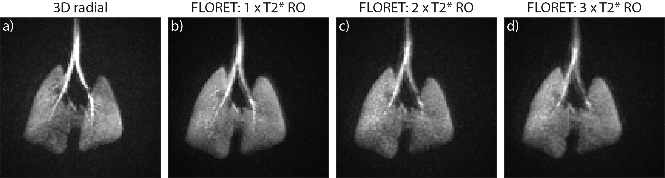
Figure 2. Representative max
intensity projections in the coronal plane from one mouse. a) 3D radial, 25%
percent sampled. b-d) 3D spiral (FLORET), with each spiral arm durations equal
to b) 1 x T2* readout, c) 2 x T2* readout, and d) 3 x T2* readout.
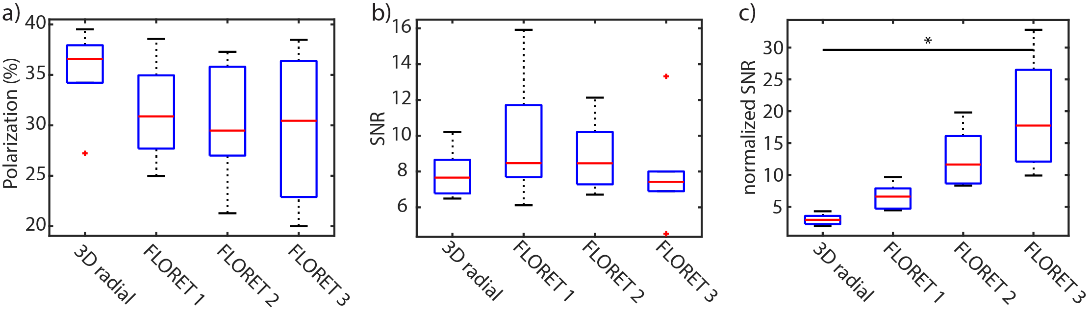
Figure 4. Box plots summarize
the values of a) estimated initial polarization, b) Signal-to-noise ratio, and
c) the corrected signal-to-noise ratio for each of the four scans. Descriptions
for the calculation of SNR and the corrected SNR are detailed in the methods
section. * ≡ significant difference (p = 0.0008).