Aurelien Bustin1,2,3, Soumaya Sridi2, Solenn Toupin4, Jerome Yerly3,5, Davide Piccini3,6, Ruud B van Heeswijk3, Pierre Jaïs1,7, Hubert Cochet1,2, and Matthias Stuber1,3,5
1IHU LIRYC, Electrophysiology and Heart Modeling Institute, Université de Bordeaux, INSERM, Centre de recherche Cardio-Thoracique de Bordeaux, U1045, Bordeaux, France, 2Department of Cardiovascular Imaging, Hôpital Cardiologique du Haut-Lévêque, CHU de Bordeaux, Bordeaux, France, 3Department of Diagnostic and Interventional Radiology, Lausanne University Hospital and University of Lausanne, Lausanne, Switzerland, 4Siemens Healthcare France, Saint-Denis, France, 5Center for Biomedical Imaging (CIBM), Lausanne, Switzerland, 6Advanced Clinical Imaging Technology, Siemens Healthcare, Lausanne, Switzerland, 7Department of Cardiac Electrophysiology, Hôpital Cardiologique du Haut-Lévêque, CHU de Bordeaux, Bordeaux, France
1IHU LIRYC, Electrophysiology and Heart Modeling Institute, Université de Bordeaux, INSERM, Centre de recherche Cardio-Thoracique de Bordeaux, U1045, Bordeaux, France, 2Department of Cardiovascular Imaging, Hôpital Cardiologique du Haut-Lévêque, CHU de Bordeaux, Bordeaux, France, 3Department of Diagnostic and Interventional Radiology, Lausanne University Hospital and University of Lausanne, Lausanne, Switzerland, 4Siemens Healthcare France, Saint-Denis, France, 5Center for Biomedical Imaging (CIBM), Lausanne, Switzerland, 6Advanced Clinical Imaging Technology, Siemens Healthcare, Lausanne, Switzerland, 7Department of Cardiac Electrophysiology, Hôpital Cardiologique du Haut-Lévêque, CHU de Bordeaux, Bordeaux, France
Myocardial T1 rho mapping with model-based non-rigid motion correction enables quantitative characterization of myocardial injuries with relatively low sensitivity to respiratory motion and field inhomogeneity.
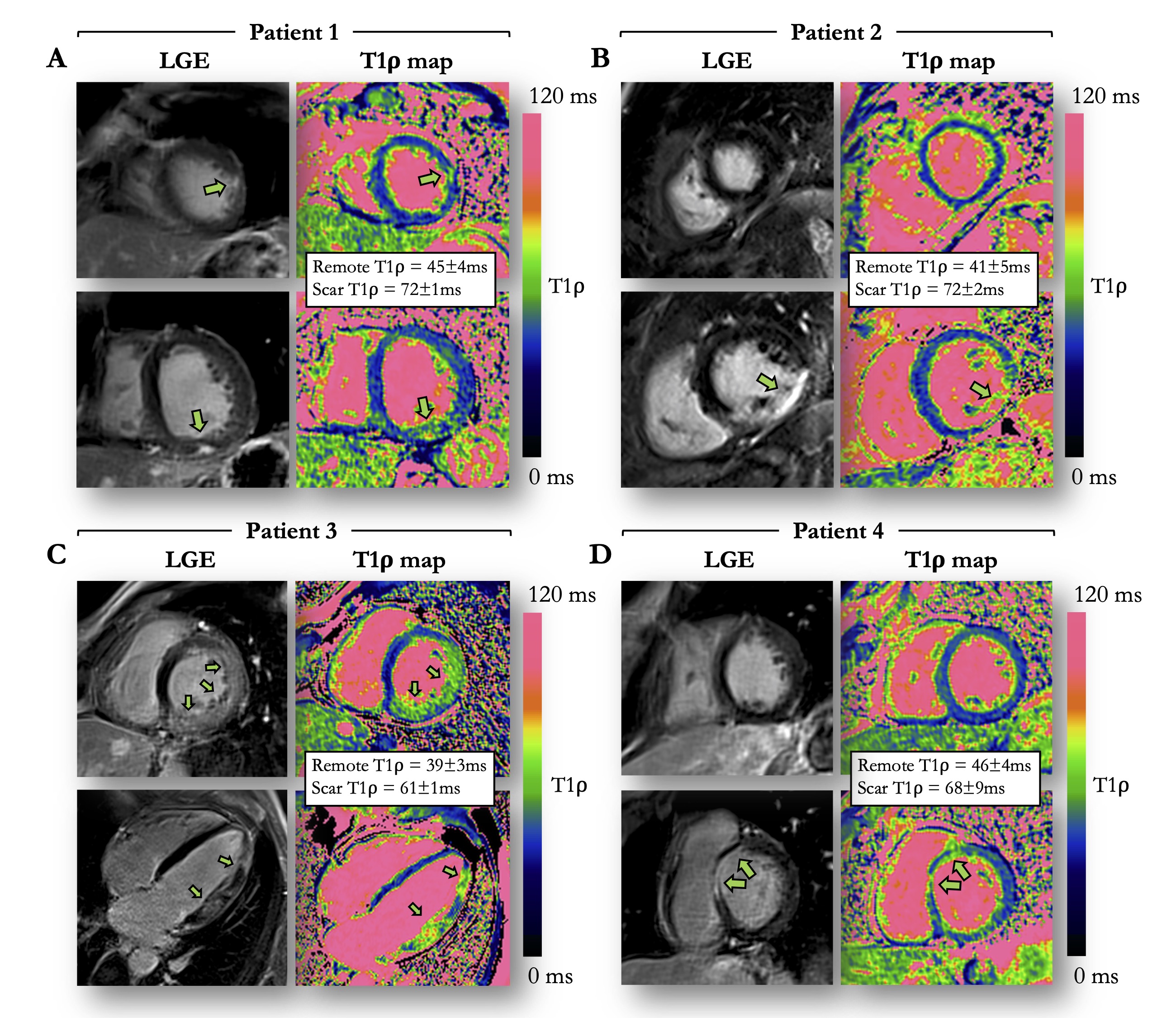
Figure 3: Examples of 4 patients with evidence of myocardial injury on LGE and motion-corrected T1ρ mapping. (A) 59-year-old male patient with sub-epicardial LGE in the latero-apical segment. (B) 53-year-old male patient with ischemic cardiomyopathy and transmural LGE in the inferior and infero-septal mid segments. (C) 51-year-old male patient with acute myocarditis and extensive patchy intramural and subepicardial LGE in the left ventricular free wall. (D) 35-year-old male patient with myocarditis and intramural LGE in the antero-septo-basal segment.
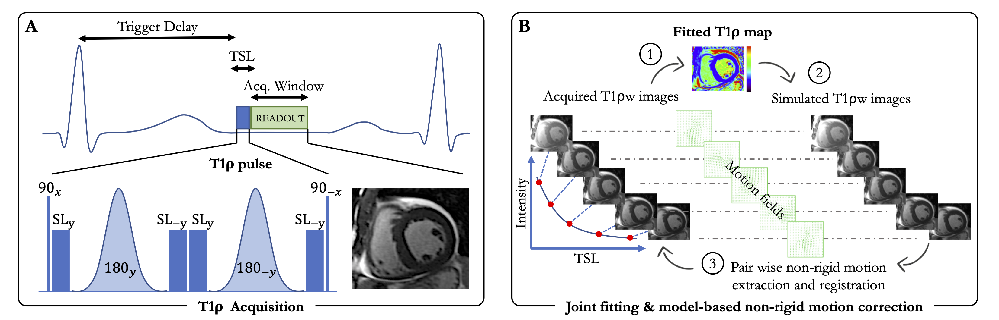
Figure 1: Schematic of the proposed single breath-hold 2D myocardial T1ρ mapping technique (A) with joint T1ρ fitting and model-based motion correction (B). T1ρ mapping is performed using a single-shot electrocardiogram-triggered bSSFP acquisition where five images with different spin lock times are acquired within a single breath-hold. Motion correction is performed by iterating between a T1ρ fitting (step 1), the simulation of T1ρ-weighted images (step 2) and a pair-wise non-rigid motion correction (step 3).