Xianglun Mao1, Fardad M Serry1, Sen Ma1, Zhehao Hu1,2, Alan C Kwan1,3, Fei Han4, Yibin Xie1, Debiao Li1,2, and Anthony G Christodoulou1,2
1Biomedical Imaging Research Institute, Cedars-Sinai Medical Center, Los Angeles, CA, United States, 2Department of Bioengineering, University of California in Los Angeles, Los Angeles, CA, United States, 3Smidt Heart Institute, Cedars-Sinai Medical Center, Los Angeles, CA, United States, 4Siemens Medical Solutions Inc., Los Angeles, CA, United States
1Biomedical Imaging Research Institute, Cedars-Sinai Medical Center, Los Angeles, CA, United States, 2Department of Bioengineering, University of California in Los Angeles, Los Angeles, CA, United States, 3Smidt Heart Institute, Cedars-Sinai Medical Center, Los Angeles, CA, United States, 4Siemens Medical Solutions Inc., Los Angeles, CA, United States
The 3D Multitasking acquisition technique simultaneously acquires co-registered T1, T2, and B1+ maps with cine capability in whole ventricle. Multitasking resolves the cardiac and respiratory motions, allowing for free-breathing acquisition without need for ECG.
Fig.5 (animated): Cardiac motion-resolved images of two subjects of proposed 3D Stack-of-Stars Multitasking in short axis orientation with 3D (1.4x1.4x8.0 mm3 resolution) whole-ventricle coverage in TA=9:14min. Comparative multi-slice and multi breath-hold 2D TrueFisp CINE in short axis (non-fat suppressed, 1.3x1.3x8.0 mm3 resolution, TA=11s per slice).
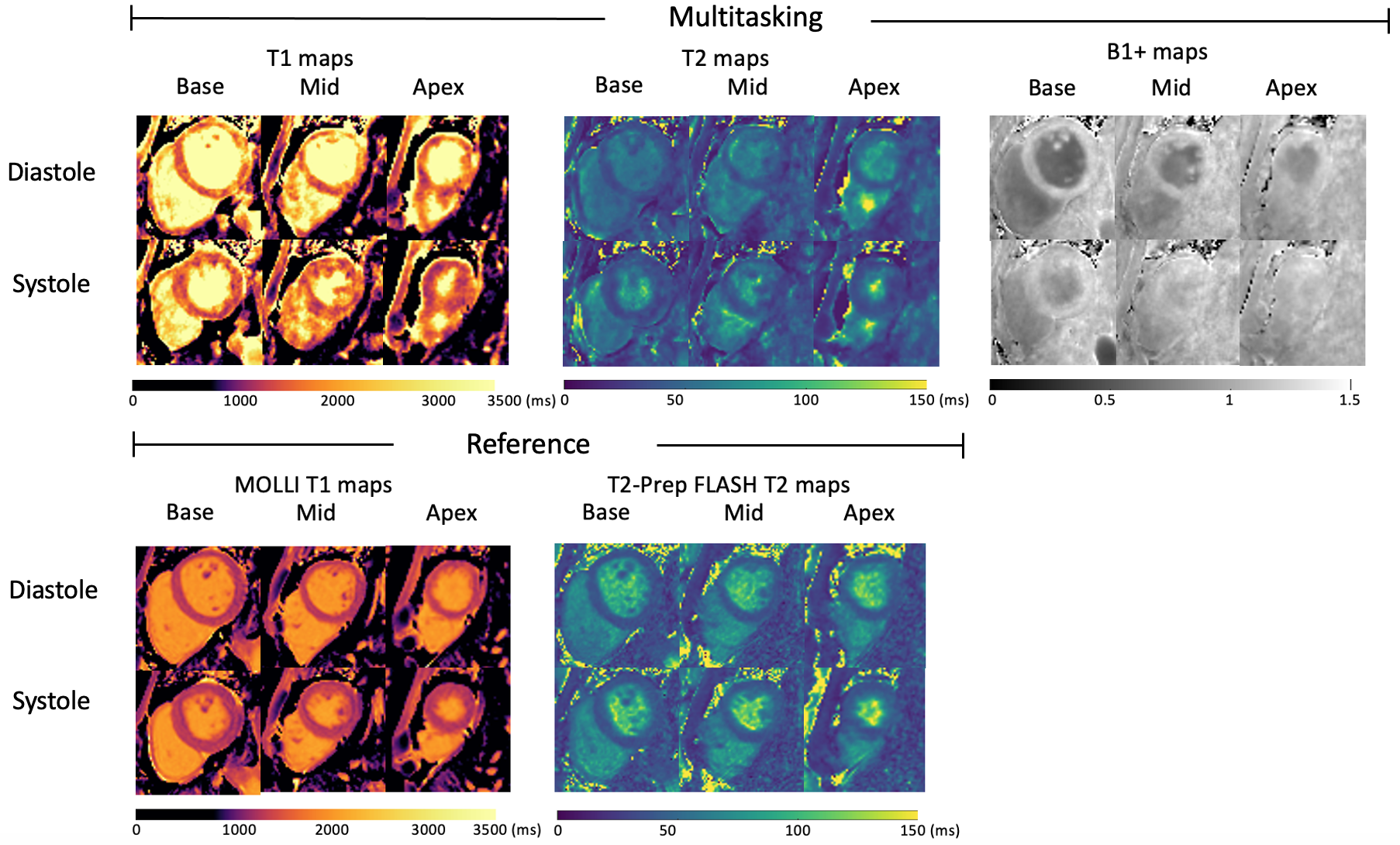
Fig.3: The proposed 3D Multitasking T1,T2, B1+ cine mapping results (basal, mid and apical) and short-axis 2D MOLLI T1 maps, T2-prep FLASH T2 maps for one subject (F, 27).