Carlos Velasco1, Gastao Cruz1, René M. Botnar1, and Claudia Prieto1
1School of Biomedical Engineering and Imaging Sciences, King's College London, London, United Kingdom
1School of Biomedical Engineering and Imaging Sciences, King's College London, London, United Kingdom
A cardiac MRF acquisition scheme for simultaneous quantification of myocardial T1, T2 and T1ρ in a contrast-free single breath-hold MR scan of ~16s is proposed.
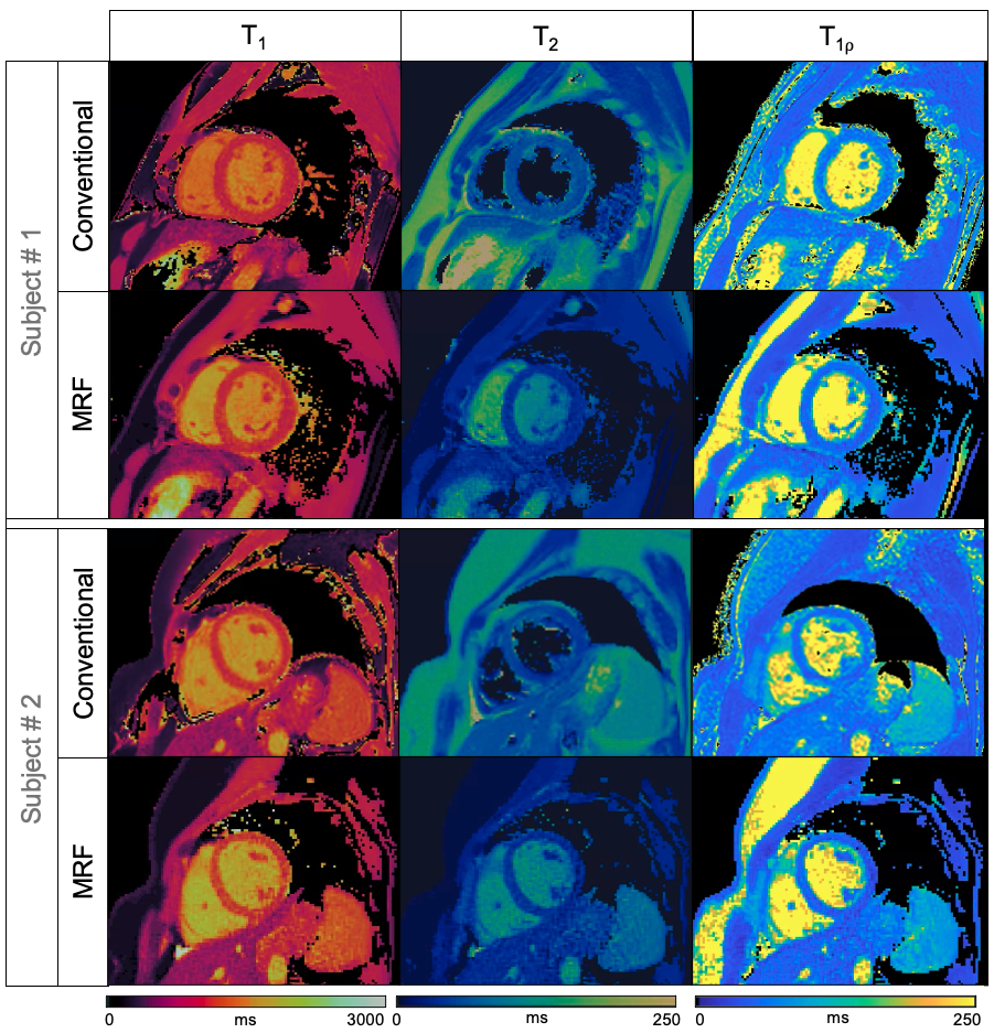
Fig 4: Short axis view of T1, T2 and T1ρ quantitative maps in two representative healthy subjects. Reference maps (top row) of T1-MOLLI, T2-GRaSE and T1⍴-Reference are compared against the regularized T1, T2 and T1⍴ cardiac MRF maps (bottom row) obtained from the same ~16s cardiac MRF acquisition.
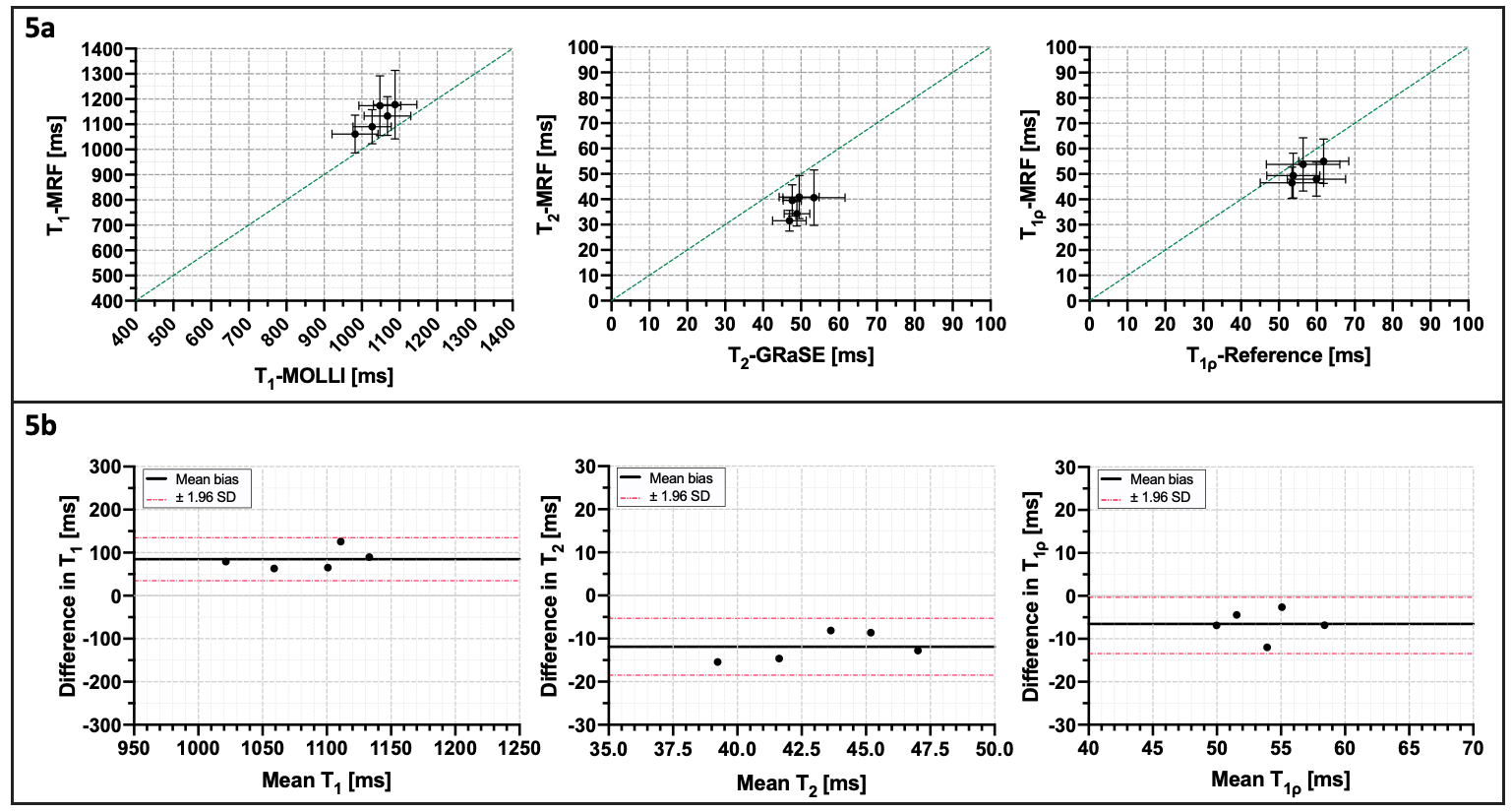
Fig 5: a) Scatter plots and T1, T2 and T1ρ cardiac MRF quantification compared against their reference values obtained from T1-MOLLI, T2-GRaSE and T1⍴-Reference. Green dashed lines depict the identity line. b) Bland-Altman plots of T1, T2 and T1ρ cardiac MRF against their respective references. Black lines denote the mean bias and dashed-dotted red lines show the limits of agreement (95% confidence interval).