Simone Rumac1, Christopher W. Roy2, Jérôme Yerly2,3, John Heerfordt2,4, Davide Piccini2,4, Matthias Stuber3,5, and Ruud B. van Heeswijk2
1Department of Radiology, Department of Radiology, Lausanne University Hospital (CHUV) and University of Lausanne (UNIL), Laus, Lausanne, Switzerland, 2Department of Radiology, Department of Radiology, Lausanne University Hospital (CHUV) and University of Lausanne (UNIL), Lausanne, Switzerland, Lausanne, Switzerland, 3CIBM Center for BioMedical Imaging, Lausanne, Switzerland, Lausanne, Switzerland, 4Advanced Clinical Imaging Technology, Siemens Healthcare AG, Lausanne, Switzerland, Lausanne, Switzerland, 5Department of Radiology, Department of Radiology, Lausanne University Hospital (CHUV) and University of Lausanne, Lausanne, Switzerland
1Department of Radiology, Department of Radiology, Lausanne University Hospital (CHUV) and University of Lausanne (UNIL), Laus, Lausanne, Switzerland, 2Department of Radiology, Department of Radiology, Lausanne University Hospital (CHUV) and University of Lausanne (UNIL), Lausanne, Switzerland, Lausanne, Switzerland, 3CIBM Center for BioMedical Imaging, Lausanne, Switzerland, Lausanne, Switzerland, 4Advanced Clinical Imaging Technology, Siemens Healthcare AG, Lausanne, Switzerland, Lausanne, Switzerland, 5Department of Radiology, Department of Radiology, Lausanne University Hospital (CHUV) and University of Lausanne, Lausanne, Switzerland
We
developed a novel fast high-resolution free-breathing 3D isotropic T2
mapping technique for the heart. The resulting whole-heart T2 maps
were both accurate and sharp, and the T2 values in the myocardium matched
those measured with routine 2D T2 mapping (p=0.57).
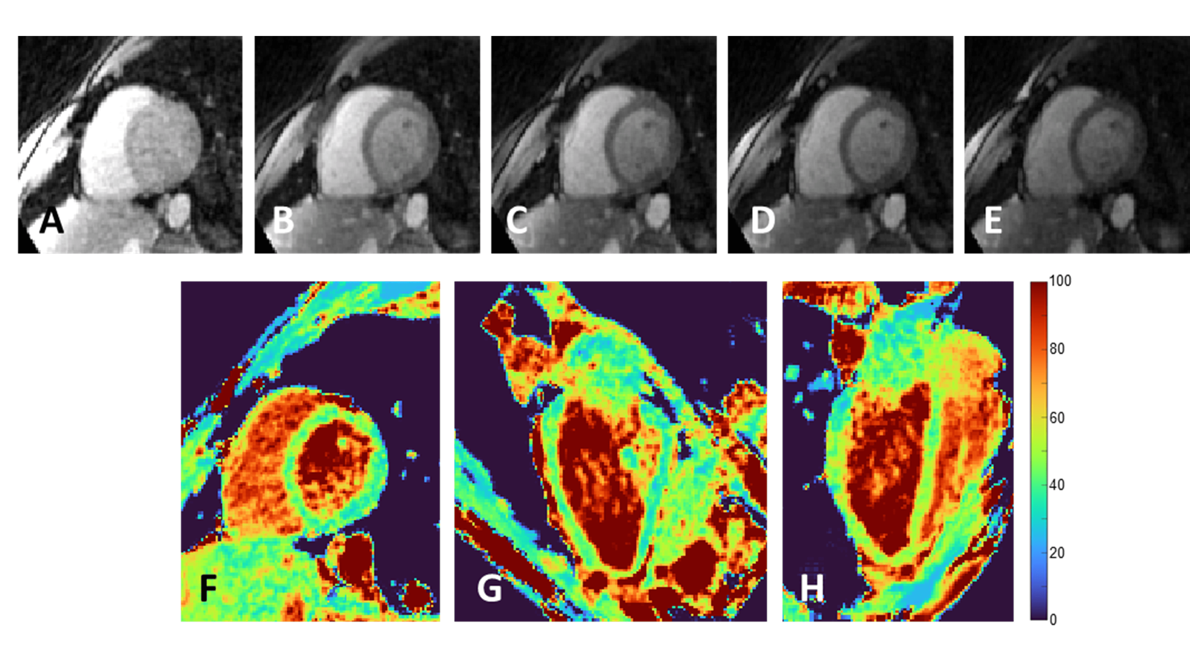
Sharp source images and precise T2 maps in healthy volunteers. A-E) The source images contain sharp
features (papillary muscles, edge definition) and are well aligned to one
another. F-H) The resulting short-axis T2 map and its two perpendicular views are
similarly sharp.
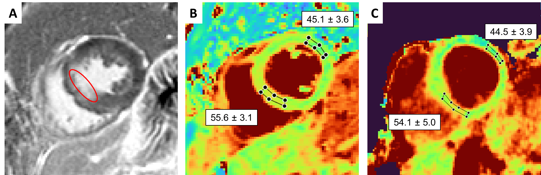
T2 mapping in a patient with myocardial infarction.
A) LGE can be observed in the septum. B) The corresponding T2
map obtained with 2D T2-prepared bSSFP shows a small T2 elevation
in the infarcted area. C) The proposed technique accurately confirms the T2
elevation.