Ruixi Zhou1, Daniel S. Weller2, Yang Yang3, Junyu Wang1, John P. Mugler4, and Michael Salerno5
1Biomedical Engineering, University of Virginia, Charlottesville, VA, United States, 2Electrical and Computer Engineering, University of Virginia, Charlottesville, VA, United States, 3Biomedical Engineering and Imaging Institute and Department of Radiology, Icahn School of Medicine at Mount Sinai, New York, NY, United States, 4Radiology, Biomedical Engineering, University of Virginia, Charlottesville, VA, United States, 5Cardiology, Radiology, Biomedical Engineering, University of Virginia, Charlottesville, VA, United States
1Biomedical Engineering, University of Virginia, Charlottesville, VA, United States, 2Electrical and Computer Engineering, University of Virginia, Charlottesville, VA, United States, 3Biomedical Engineering and Imaging Institute and Department of Radiology, Icahn School of Medicine at Mount Sinai, New York, NY, United States, 4Radiology, Biomedical Engineering, University of Virginia, Charlottesville, VA, United States, 5Cardiology, Radiology, Biomedical Engineering, University of Virginia, Charlottesville, VA, United States
In a single acquisition, a free breathing Bloch-Siegert
shift B1 map, and a self-gated B1 and slice profile corrected T1 map are
acquired. The technique is compared to our prior dual-flip angle approach and yields
more accurate T1 values.
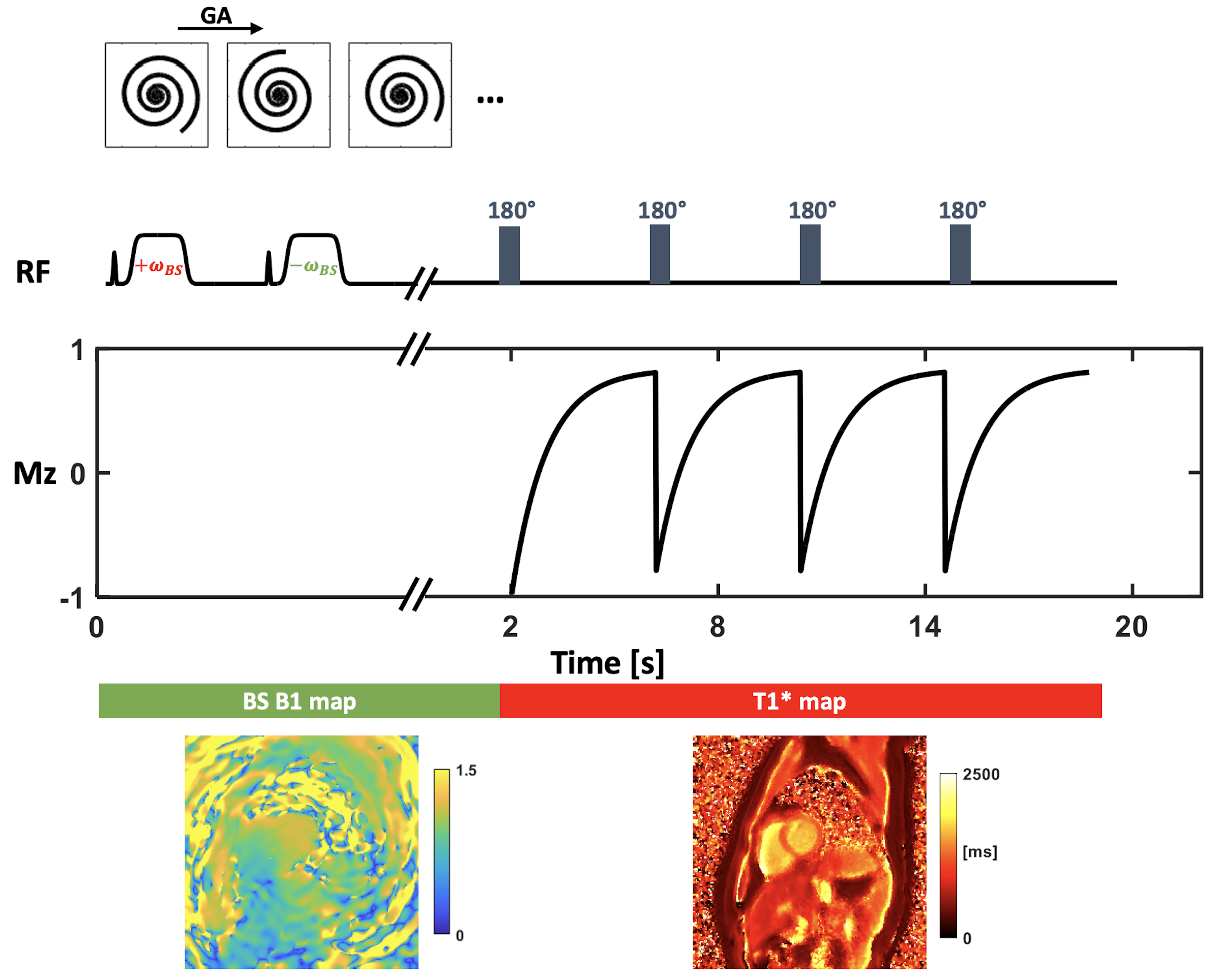
Figure 1. Schematic of the acquisition. Data in the
first two seconds acquired with off-resonance Fermi pulses were used to
reconstruct the Bloch-Siegert B1 map. Data acquired with repetitive inversion
pulses were used to reconstruct the T1* map.
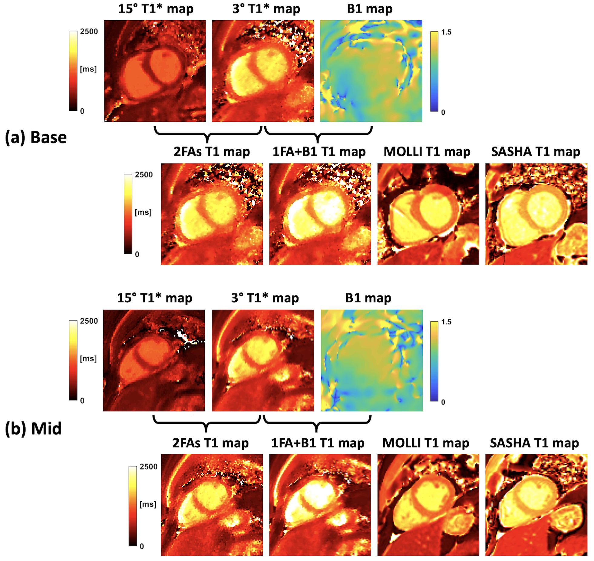
Figure 4. Human results. Short-axis basal (a) and
middle (b) slices from one human subject are shown. As indicated by the
bracket, 3° and 15° T1* maps are used to generate the 2FAs T1 map, while 3° T1*
and BS B1 maps are used to generate the 1FA+B1 T1 map. MOLLI and SASHA T1 maps
are also shown as comparison.