Dan Wu1, Kewen Jiang2, Yi-Cheng Hsu3, Yi Sun3, Yi Zhang1, and Yudong Zhang2
1Biomedical Engineering, Zhejiang University, Hangzhou, China, 2Radiology, the First Affiliated Hospital with Nanjing Medical University, Nanjing, China, 3MR Collaboration, Siemens Healthcare Ltd., Shanghai, China
1Biomedical Engineering, Zhejiang University, Hangzhou, China, 2Radiology, the First Affiliated Hospital with Nanjing Medical University, Nanjing, China, 3MR Collaboration, Siemens Healthcare Ltd., Shanghai, China
In prostate cancer, fin and cellularity obtained from diffusion time-dependent diffusion MRI
and IMPULSED model increased as Gleason score increased, while and diameter and
Dex decreased. Cellularity
achieved the highest diagnostic accuracy with an area-under-the curve of 0.96.
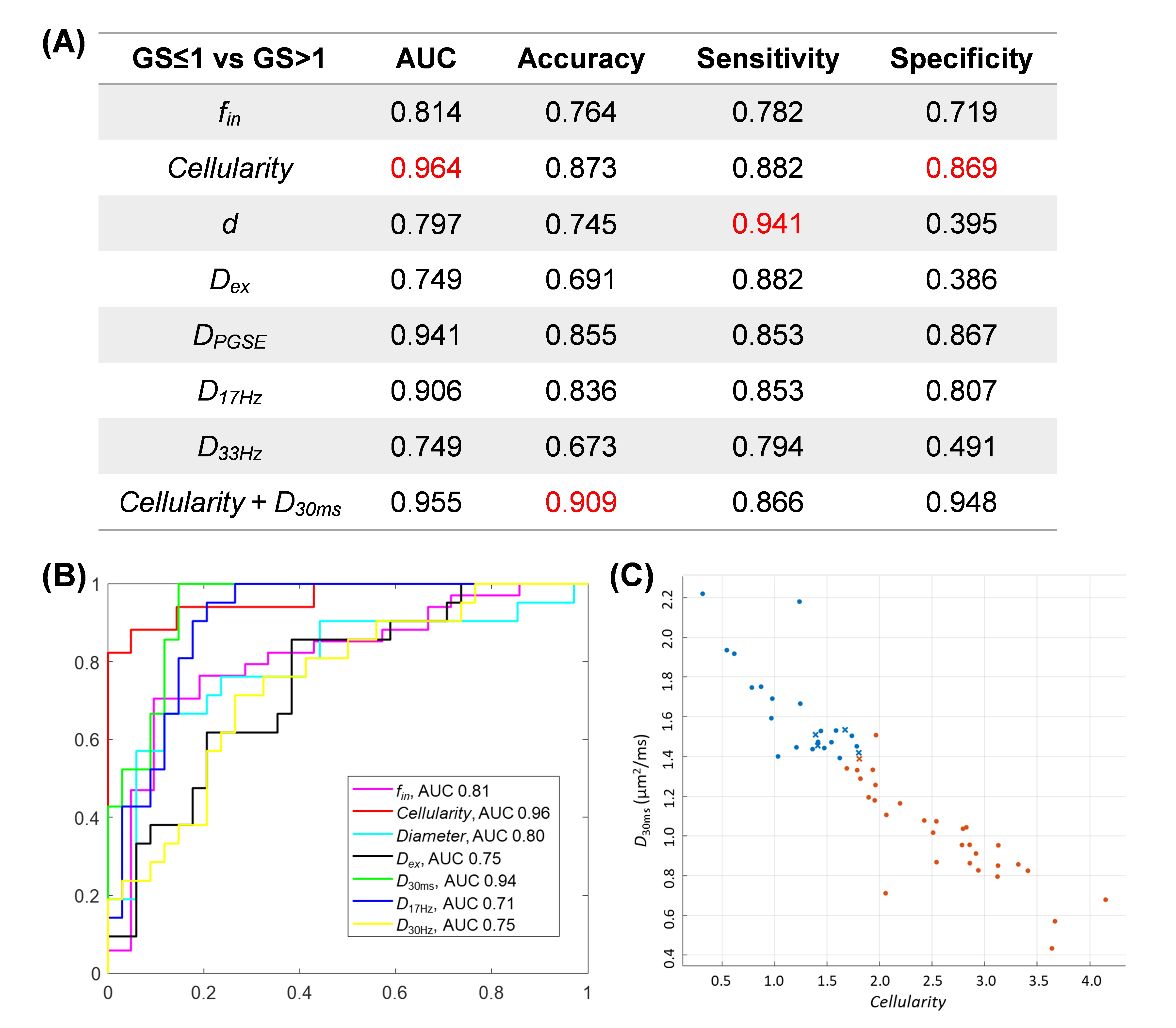
Figure 4:
(A) Diagnostic performance of different microstructural markers to differeciate
clinically significant (GS>1) and insignificant (GS≤1) cancers in terms of
area-under-the curve (AUC), accuracy, sensitivity and specificity, based on a
five-fold cross-validation using the linear discriminator. (B)
Receiver-operating-curves (ROCs) of the different markers. (C) Classifying
clinically significant (red) and insignificant (blue) cancers using the
combined marker of the cellularity and DPGSE(30ms).
The mis-classified cases are marked as “x”.
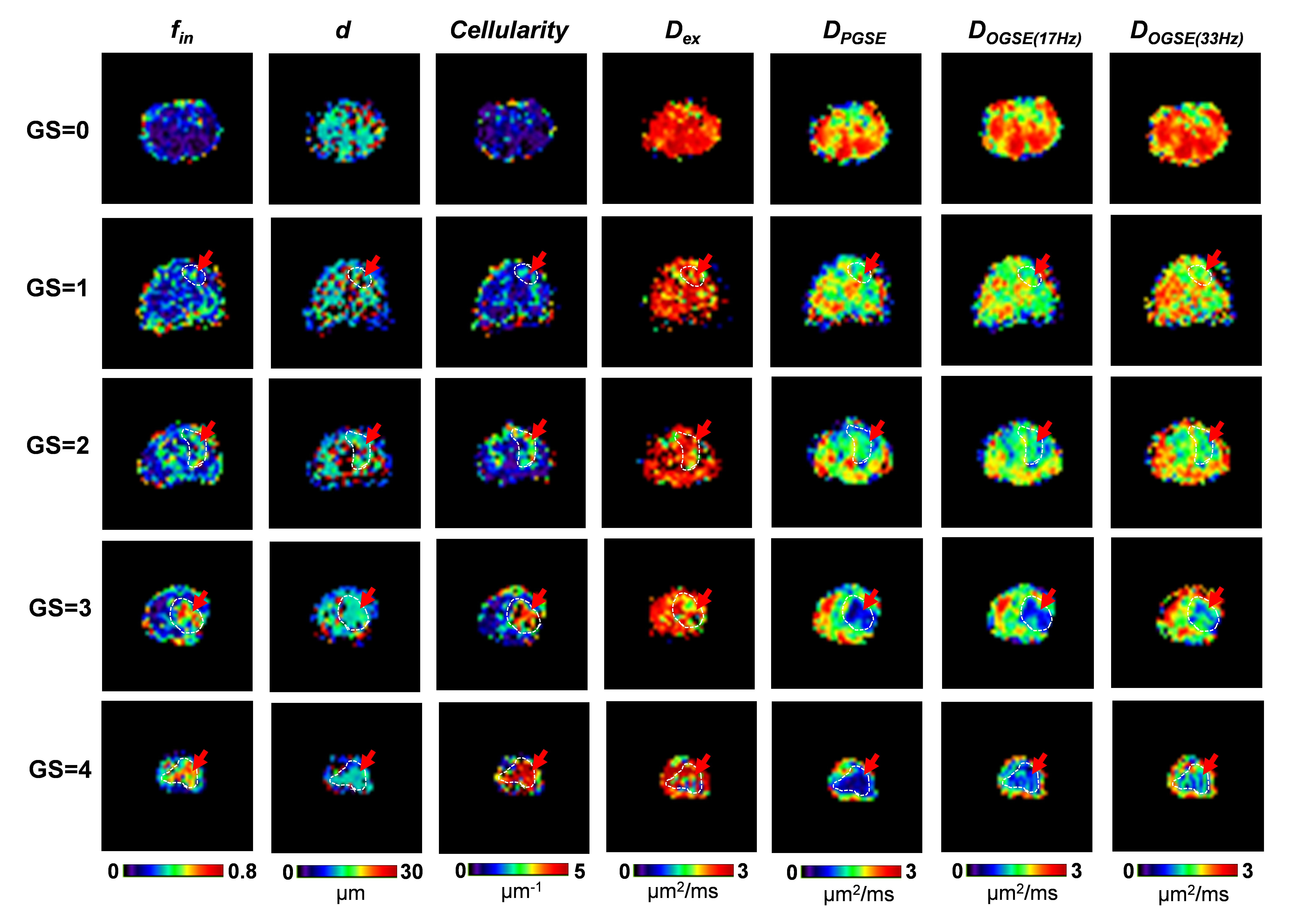
Figure 1:
Microstructural maps of the prostate tissues of Gleason score (GS) from 0-4. Intracellular
fraction (fin), cell
diameter (d), cellularity, and
extracellular diffusivity (Dex)
fitted from the IMPULSED model and the diffusivity maps from PGSE, OGSE (17Hz),
and OGSE (33Hz) data were shown. The white contours and red arrows indicated the
cancerous regions based on manual delineation.