Scott D. Swanson1, Thomas L. Chenevert1, Prasad R. Shankar1, Ted Lynch2, and Dariya I. Malyarenko1
1Department of Radiology, University of Michigan, Ann Arbor, MI, United States, 2CIRS, Norfolk, VA, United States
1Department of Radiology, University of Michigan, Ann Arbor, MI, United States, 2CIRS, Norfolk, VA, United States
A biomimetic phantom has been developed with a range of multi-compartment diffusion parameters similar to those observed in cancerous and health prostate tissue.
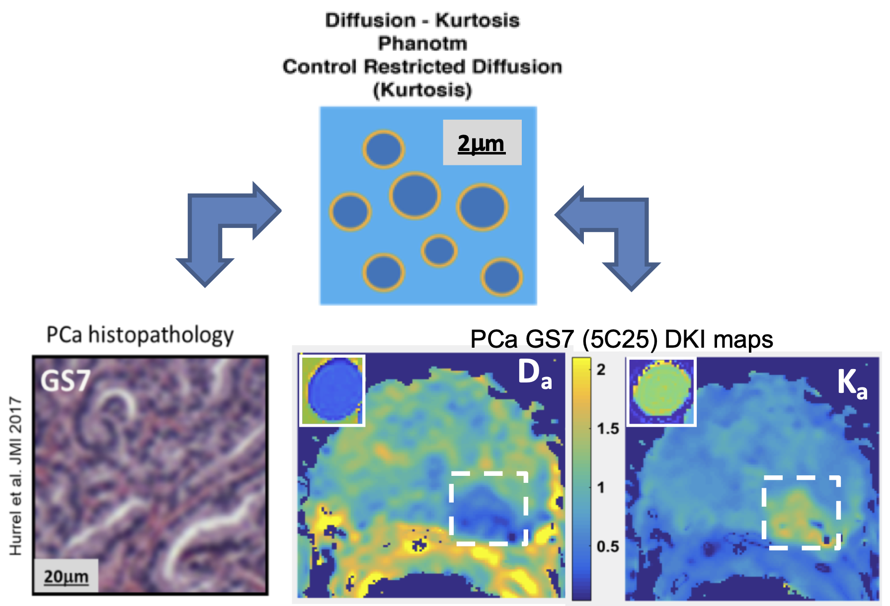
Figure 1. Schematic of biomimetic lipid nanoparticle phantom to reflect multi-compartment contribution to DWI voxel signal in PCa (GS7 histology) and example fit parametric maps for diffusion-kurtosis model. Sizes of the lipid nanoparticles can be controlled to be between 200 and 2,000 nm, depending on the composition of the nanoparticle shell.
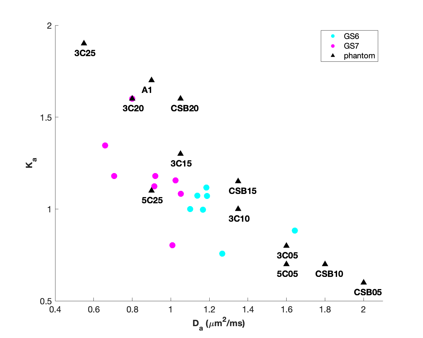
Figure 3. Scatter plot of apparent kurtosis, Ka, versus apparent diffusion, Da, for in vivio PCa and selected diffusion-kurtosis phantom materials. The range of values found in PCa GS6 and GS7 lesions for Da and Ka (estimated by fitting DWI decay curves to the isotropic diffusion kurtosis model) can be spanned by fabrication of lipid nano-materials with appropriate physical properties.