Francesco Grussu1, Ignasi Barba2, Kinga Bernatowicz1, Irene Casanova-Salas3, Alba Escriche Villarroya4, Natalia Castro3, Emanuela Greco4, Juan Francisco Corral5,6, Marta Vidorreta7, Manuel Escobar Amores5,6, Núria Roson5,6, Xavier Merino5,6, Richard Mast5,6, Nahúm Calvo‐Malvar5,8, Joaquin Mateo3, Paolo Nuciforo9, María Abad4, Josep R. Garcia-Bennett8, and Raquel Perez-Lopez1,6
1Radiomics Group, Vall d'Hebron Institute of Oncology, Vall d'Hebron Barcelona Hospital Campus, Barcelona, Spain, 2NMR Lab, Vall d'Hebron Institute of Oncology, Vall d'Hebron Barcelona Hospital Campus, Barcelona, Spain, 3Prostate Cancer Translational Research Group, Vall d'Hebron Institute of Oncology, Vall d'Hebron Barcelona Hospital Campus, Barcelona, Spain, 4Cellular Plasticity and Cancer Group, Vall d'Hebron Institute of Oncology, Vall d'Hebron Barcelona Hospital Campus, Barcelona, Spain, 5IDI (Institut de Diagnòstic per la Imatge), Catalonia, Spain, 6Department of Radiology, Hospital Universitari Vall d'Hebron, Barcelona, Spain, 7Siemens Healthineers, Madrid, Spain, 8Hospital Universitari de Bellvitge, L'Hospitalet de Llobregat, Spain, 9Molecular Oncology Group, Vall d'Hebron Institute of Oncology, Vall d'Hebron Barcelona Hospital Campus, Barcelona, Spain
1Radiomics Group, Vall d'Hebron Institute of Oncology, Vall d'Hebron Barcelona Hospital Campus, Barcelona, Spain, 2NMR Lab, Vall d'Hebron Institute of Oncology, Vall d'Hebron Barcelona Hospital Campus, Barcelona, Spain, 3Prostate Cancer Translational Research Group, Vall d'Hebron Institute of Oncology, Vall d'Hebron Barcelona Hospital Campus, Barcelona, Spain, 4Cellular Plasticity and Cancer Group, Vall d'Hebron Institute of Oncology, Vall d'Hebron Barcelona Hospital Campus, Barcelona, Spain, 5IDI (Institut de Diagnòstic per la Imatge), Catalonia, Spain, 6Department of Radiology, Hospital Universitari Vall d'Hebron, Barcelona, Spain, 7Siemens Healthineers, Madrid, Spain, 8Hospital Universitari de Bellvitge, L'Hospitalet de Llobregat, Spain, 9Molecular Oncology Group, Vall d'Hebron Institute of Oncology, Vall d'Hebron Barcelona Hospital Campus, Barcelona, Spain
We introduce DR-HIGADOS, a new liver diffusion-relaxation MRI technique
providing indices of cell size and cellularity. DR-HIGADOS is shown to be
feasible on multi-vendor clinical scans, and preliminary MRI-histology
correlations confirm its biological specificity.
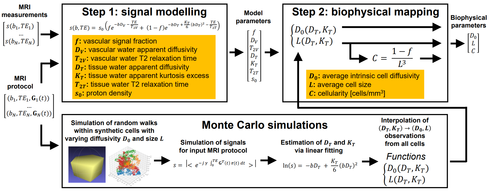
Figure
1: overview of the two-step DR-HIGADOS framework. In the
first step, an extension of the IVIM model is fitted to diffusion-relaxation
measurements. In the second step, model parameters are mapped to biophysical
properties (e.g. average intrinsic cell diffusivity and size; cellularity),
using information derived from Monte Carlo simulations.
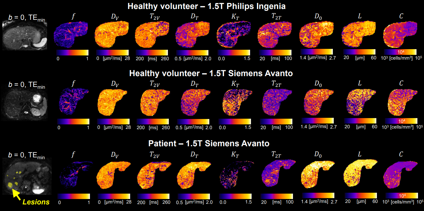
Figure 2: parametric maps provided by DR-HIGADOS in vivo
on two MRI vendors. Left to right: non-DW image, vascular
signal fraction
f
, vascular diffusivity and T2 (Dv and
T2V), tissue diffusivity, kurtosis excess and T2 (DT, KT and
T2T), average cell diffusivity and size (D0 and
L), cellularity
C. Top to bottom: healthy volunteer on Ingenia system;
healthy volunteer on Avanto system; patient on Avanto system.