Carlos Bilreiro1,2, Francisca F Fernandes1, Rui V Simões1, Mireia Castillo-Martin1,3, Andrada Ianus1, Cristina Chavarrias1, Celso Matos1,2, and Noam Shemesh1
1Champalimaud Research, Champalimaud Centre for the Unknown, Lisbon, Portugal, 2Department of Radiology, Champalimaud Clinical Centre, Lisbon, Portugal, 3Department of Pathology, Champalimaud Clinical Centre, Lisbon, Portugal
1Champalimaud Research, Champalimaud Centre for the Unknown, Lisbon, Portugal, 2Department of Radiology, Champalimaud Clinical Centre, Lisbon, Portugal, 3Department of Pathology, Champalimaud Clinical Centre, Lisbon, Portugal
We developed
a diffusion-MRI (dMRI) approach for mapping pancreatic cancer precursor lesions
in transgenic mice via ex vivo dMRI Microscopy with histological
validation, and applied the methods in vivo. Highly sensitive contrasts
were discovered.
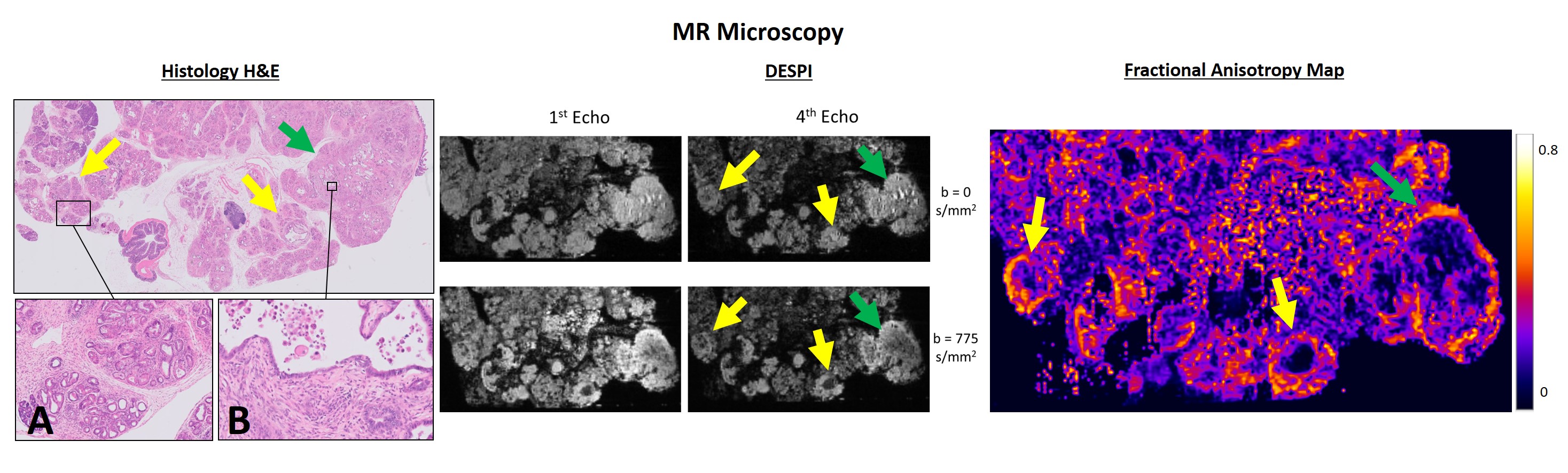
MR Microscopy, Pdx1-Cre;KrasG12D
mouse pancreas. A – PanIN in ADM background. B – Area of PDAC. As observed in
the previous example, PanIN (yellow arrows) are most conspicuous with high b
values and longer echoes, with high peripheral anisotropy values. PDAC (green
arrows) presents with similar contrasts, but with lower anisotropy values in
its solid central portions, possibly due to a different cellular and fibrotic
content.
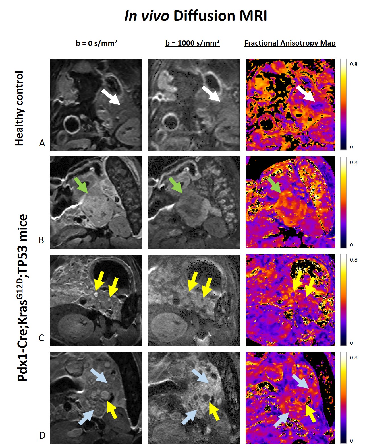
In vivo dMRI. (A) The healthy
pancreas has low signal intensity in DWI and low anisotropy values. (B) A large
abdominal mass is seen, compatible with PDAC (green arrows), with high DWI
signal intensity and high anisotropy values in its solid areas. (C) Much
smaller lesions, compatible with PanIN (yellow arrows), are depicted with
similar contrasts. (D) Similar lesions are observed (yellow arrows), along with
diffuse pancreatic changes compatible with PanIN/ADM (blue arrows).