Sungheon Gene Kim1, Mehran Baboli1, Justin Fogarty2, Steven H. Baete2, Joseph Kim3, Paulina Galavis3, Moses Tam3, Kenneth Hu3, and Elcin Zan2
1Radiology, Weill Cornell Medical College, New York, NY, United States, 2Radiology, New York University School of Medicine, New York, NY, United States, 3Radiation Oncology, New York University School of Medicine, New York, NY, United States
1Radiology, Weill Cornell Medical College, New York, NY, United States, 2Radiology, New York University School of Medicine, New York, NY, United States, 3Radiation Oncology, New York University School of Medicine, New York, NY, United States
Diffusion MRI at long diffusion times (200-700 ms)
found that the patients with less than
40% nodal volume shrinkage had significantly higher diffusivity at pretreatment
and lower kurtosis at week4 than the other patients with better response.
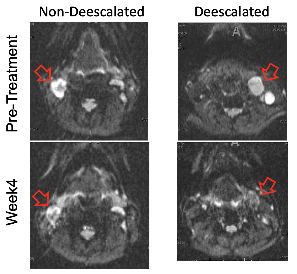
Figure 1: Example b=0 images of a non-deescalated and
deescalated patients at pre-treatment and week 4 into the treatment. The
metastatic lymph nodes are noted by arrows.
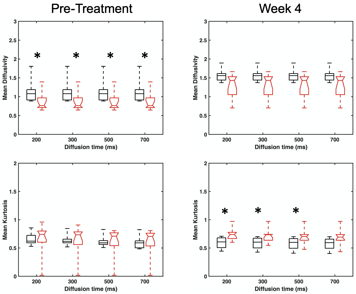
Figure 3: Box-whisker
plots of diffusivity and diffusion kurtosis of metastatic lymph nodes measured
at long diffusion times. Black square boxes are for non-deescalated patients
(n=6) and red boxes with notches are for deescalated patients (n=12).