Neele S Dellschaft1, Rachel Allcock1, Jana Hutter2, Lopa Leach3, Nia Jones4, and Penny Gowland1
1Sir Peter Mansfield Imaging Centre, University of Nottingham, Nottingham, United Kingdom, 2Department of Perinatal Imaging and Health, King's College London, London, United Kingdom, 3Life Sciences, University of Nottingham, Nottingham, United Kingdom, 4Division of Child Health, Obstetrics and Gynaecology, University of Nottingham, Nottingham, United Kingdom
1Sir Peter Mansfield Imaging Centre, University of Nottingham, Nottingham, United Kingdom, 2Department of Perinatal Imaging and Health, King's College London, London, United Kingdom, 3Life Sciences, University of Nottingham, Nottingham, United Kingdom, 4Division of Child Health, Obstetrics and Gynaecology, University of Nottingham, Nottingham, United Kingdom
Observed on T2*-weighted scans, contractions with (placental pump) and
without (Braxton-Hicks) reduction in placental volume share many
characteristics. A reduction in intensity is preceded by a change in placental
shape, with thickening of the underlying uterine wall in half the cases.
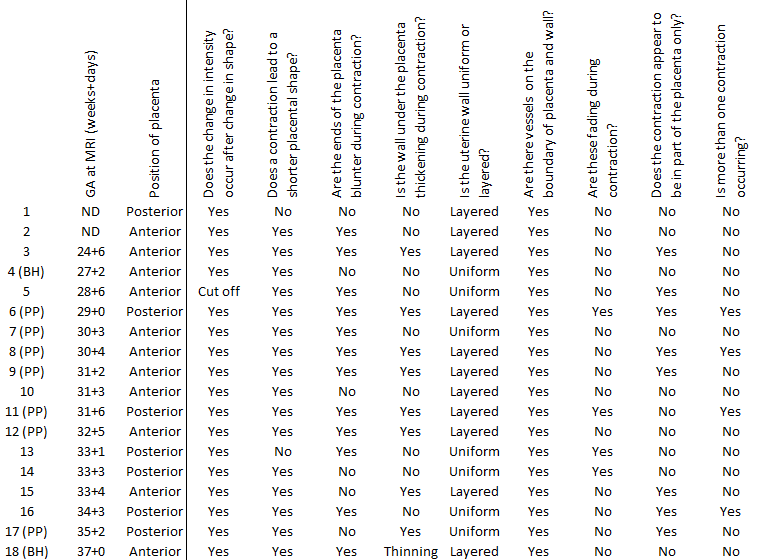
Fig
1:
Summary of findings in contractions, sorted by ascending gestational age
at scan (ND, no data for gestational age). In cases where placental volumes have
currently been measured, contractions were defined as placental pump, PP, if
contraction was accompanied by a reduction in placental volume, Braxton-Hicks,
BH, if there was no change. In subject 5, changes at the start of the
contraction could not be assessed because they had already occurred at the
start of the scan (‘cut off’).
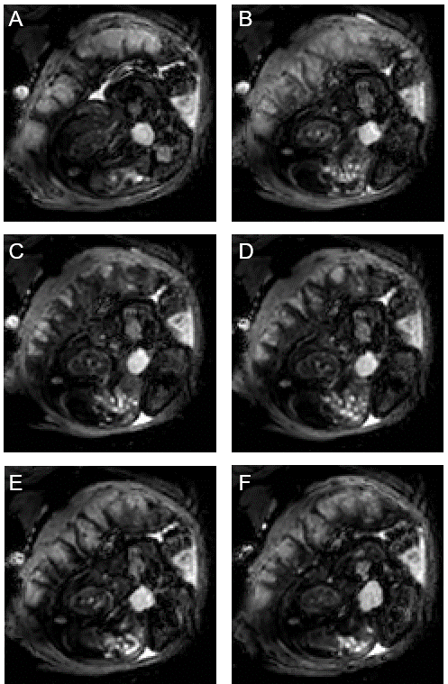
Fig 5: Typical
progression of hypointense signal during a contraction. A) before the
contraction, B) shape has started changing, thin dark lines across placenta (37
seconds after A), C) placenta is fully contracted, almost completely dark
besides some area near basal plate (72 s after A), D) dark pattern remains
longest on side of chorionic plate (82 s after A), E) pattern has faded except
for thin lines outlining cotyledons (118 s after A), F) returned to relaxed
state (179 s after A).