Datta Singh Goolaub1,2, Jiawei Xu3, Eric Schrauben4, Davide Marini5, Mike Seed5,6, and Christopher Macgowan1,2
1Medical Biophysics, University of Toronto, Toronto, ON, Canada, 2Translational Medicine, The Hospital for Sick Children, Toronto, ON, Canada, 3Labatt Family Heart Centre, The Hospital for Sick Children, Toronto, ON, Canada, 4Radiology & Nuclear Medicine, Amsterdam University Medical Centers, Amsterdam, Netherlands, 5Pediatric Cardiology, The Hospital for Sick Children, Toronto, ON, Canada, 6Department of Pediatrics, University of Toronto, Toronto, ON, Canada
1Medical Biophysics, University of Toronto, Toronto, ON, Canada, 2Translational Medicine, The Hospital for Sick Children, Toronto, ON, Canada, 3Labatt Family Heart Centre, The Hospital for Sick Children, Toronto, ON, Canada, 4Radiology & Nuclear Medicine, Amsterdam University Medical Centers, Amsterdam, Netherlands, 5Pediatric Cardiology, The Hospital for Sick Children, Toronto, ON, Canada, 6Department of Pediatrics, University of Toronto, Toronto, ON, Canada
4D flow of the fetal cardiac
vasculature using PCMRI is developed and shown to agree with conventional
imaging. Detailed visualization of complex fetal hemodynamics is presented.
Tracking
inferior vena cava (IVC) and ductus venosus (DV) blood flow across the fetal
heart. (A1) Dynamic coronal view. (A2-A4) Particle traces,
released from the IVC (blue) and DV (red), at different phases of the cardiac
cycle. Parallel streams from the IVC and DV enter the right atrium (A2),
with oxygenated blood from the DV preferential shunted into the left ventricle
(A3) to supply the coronary and cerebral circulations (A4). (A5)
Corresponding flow map, colored by hypothetical oxygenation.
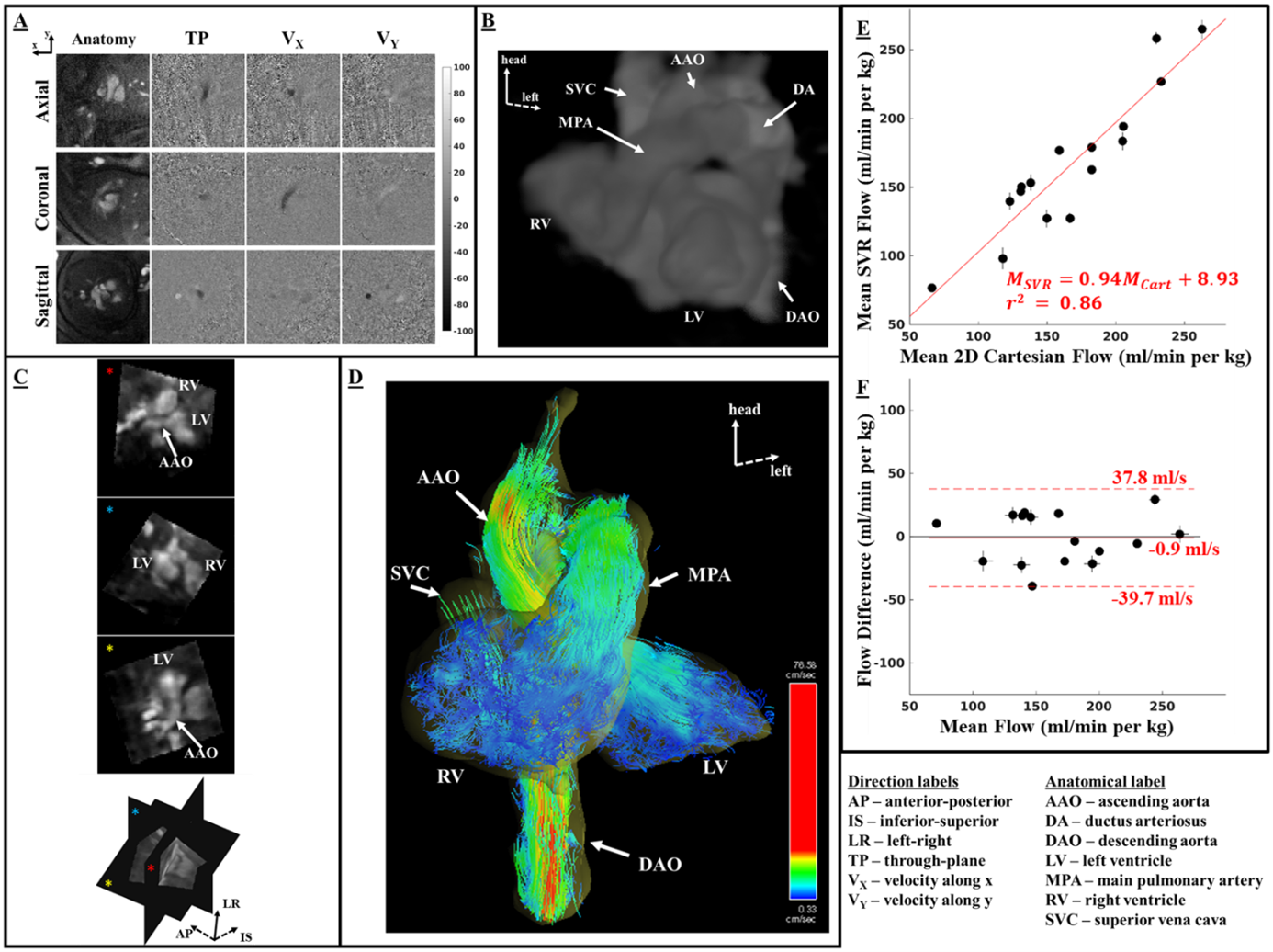
Fetal reconstructions and flow comparisons. (A)
Axial, coronal, and sagittal slices. (B) 3D rendering of slice-to-volume
reconstruction (SVR). (C) Oblique slices from reconstructed volume using
SVR. (D) Coronal view of systolic particle tracking from SVR flow.
Comparison between mean flow measurements from SVR and 2D Cartesian flow: (E)
Linear regression with red line depicting the best fit to the data and (F)
Bland-Altman plot comparing the mean flows.