Yuhao Liao1, Taotao Sun2,3, Ling Jiang2,3, Zhiyong Zhao1, Tingting Liu1, Zhaoxia Qian2,3, Yi Sun4, Yi Zhang1, and Dan Wu1
1Key Laboratory for Biomedical Engineering of Ministry of Education, Department of Biomedical Engineering, College of Biomedical Engineering & Instrument Science, Zhejiang University, Hangzhou, China, 2Department of Radiology, International Peace Maternity and Child Health Hospital, School of Medicine, Shanghai Jiao Tong University, Shanghai, China, 3Shanghai Key Laboratory of Embryo Original Diseases, Shanghai, China, 4MR Collaboration, Siemens Healthcare China, Shanghai, China
1Key Laboratory for Biomedical Engineering of Ministry of Education, Department of Biomedical Engineering, College of Biomedical Engineering & Instrument Science, Zhejiang University, Hangzhou, China, 2Department of Radiology, International Peace Maternity and Child Health Hospital, School of Medicine, Shanghai Jiao Tong University, Shanghai, China, 3Shanghai Key Laboratory of Embryo Original Diseases, Shanghai, China, 4MR Collaboration, Siemens Healthcare China, Shanghai, China
We proposed a joint analysis of flow-compensated
(FC) and non-compensated (NC) diffusion MRI to estimate the fraction
and velocity of ballistic microcirculatory flow, and evaluated the diagnostic
performance of the new IVIM markers in maternal and fetal disorders associated with placenta.
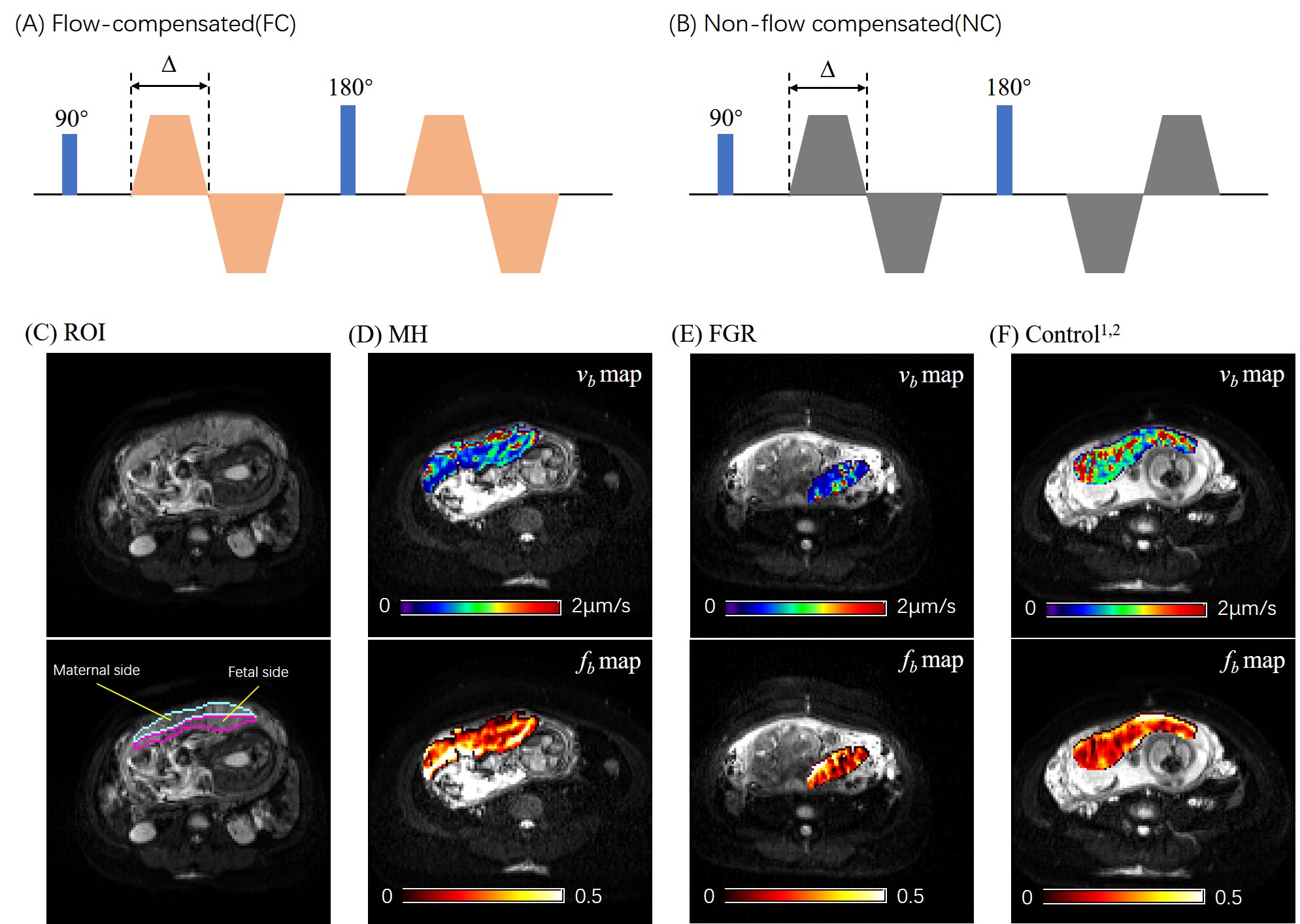
Figure 1. (A-B) Diagrams of the FC and
NC diffusion-encoding gradients with a diffusion duration of ∆. (C) Definition
of the maternal and fetal ROIs of the placenta overlaid on the b0 image. (D-F)
Representative fb and vb maps of the placenta from
a maternal hyperglycemia (MH) patient (D), a fetal growth restriction (FGR)
patient (E), and a normal control (F).
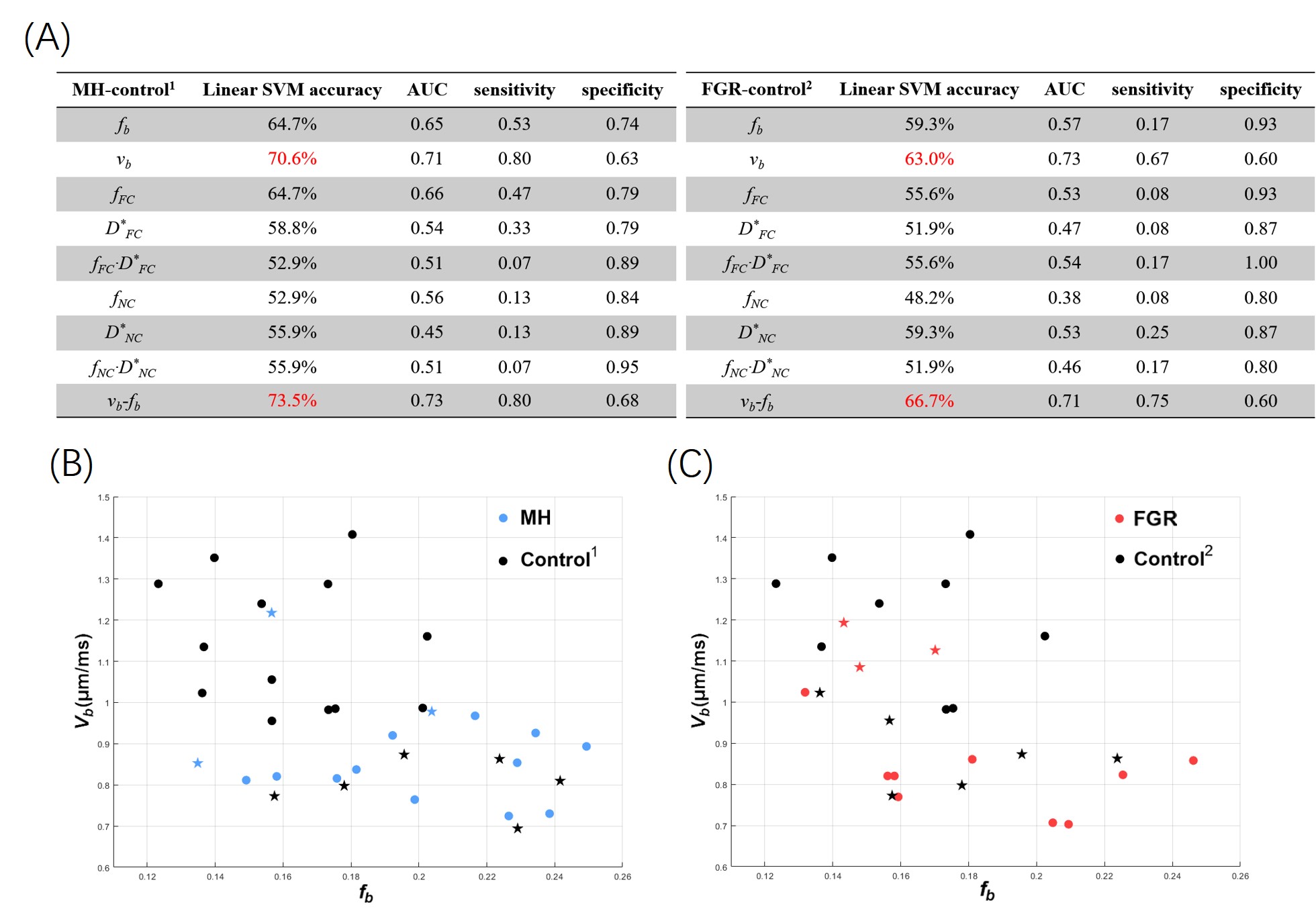
Figure 3. (A) The classification accuracy, AUC, sensitivity, and specificity of each
IVIM parameter and their combinations (as features in classification), based on
a linear SVM with five-fold cross validation. The combination of fb and vb from NC-FC model showed the best performance. (B-C) The
classification between MH and Control1 (B) and between FGR and
Control2 (C) in the two-dimensional feature space of vb-fb. The dots represent correctly identified cases, and
stars represented mis-classified cases by SVM.