Netanell Avisdris1,2, Daphna Link-Sourani2, Liat Ben-Sira3,4,5, Leo Joskowicz1, Elka Miller6, and Dafna Ben-Bashat2,3,5
1School of computer science and engineering, Hebrew University of Jerusalem, Jerusalem, Israel, 2Sagol Brain Institute, Tel Aviv Sourasky Medical Center, Tel Aviv, Israel, 3Sagol School of Neuroscience, Tel Aviv University, Tel Aviv, Israel, 4Division of Pediatric Radiology, Tel Aviv Sourasky Medical Center, Tel Aviv, Israel, 5Sackler Faculty of Medicine, Tel Aviv University, Tel Aviv, Israel, 6Medical Imaging, Children's Hospital of Eastern Ontario, University of Ottawa, Ottawa, ON, Canada
1School of computer science and engineering, Hebrew University of Jerusalem, Jerusalem, Israel, 2Sagol Brain Institute, Tel Aviv Sourasky Medical Center, Tel Aviv, Israel, 3Sagol School of Neuroscience, Tel Aviv University, Tel Aviv, Israel, 4Division of Pediatric Radiology, Tel Aviv Sourasky Medical Center, Tel Aviv, Israel, 5Sackler Faculty of Medicine, Tel Aviv University, Tel Aviv, Israel, 6Medical Imaging, Children's Hospital of Eastern Ontario, University of Ottawa, Ottawa, ON, Canada
A novel automatic method for in-vivo fetal ocular
measurements in MRI is described. Experimental results indicate that our method
matches within <1mm manual expert neuro-radiologist annotations.
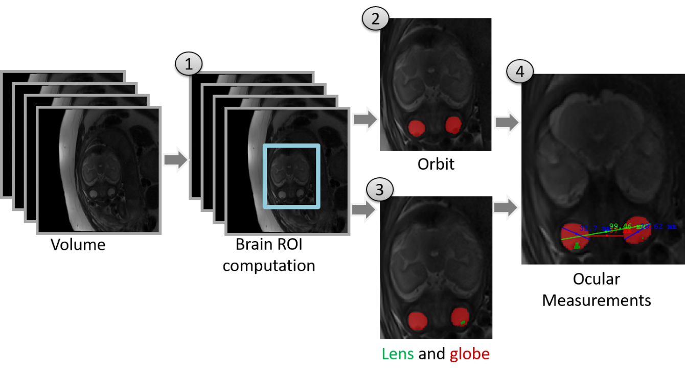
Figure 2: Illustration of the
method. The input is fetal T2 weighted volume. (1) Brain ROI
computation using 3D CNN; (2) Segmentation of the orbits for 2D
and 3D measurements using 2D U-Net; (3) Segmentation of lens and
globe for OD-LA (ocular diameter lens aligned) measurement using 2D U-Net; (4) Ocular measurements
using geometric algorithms.
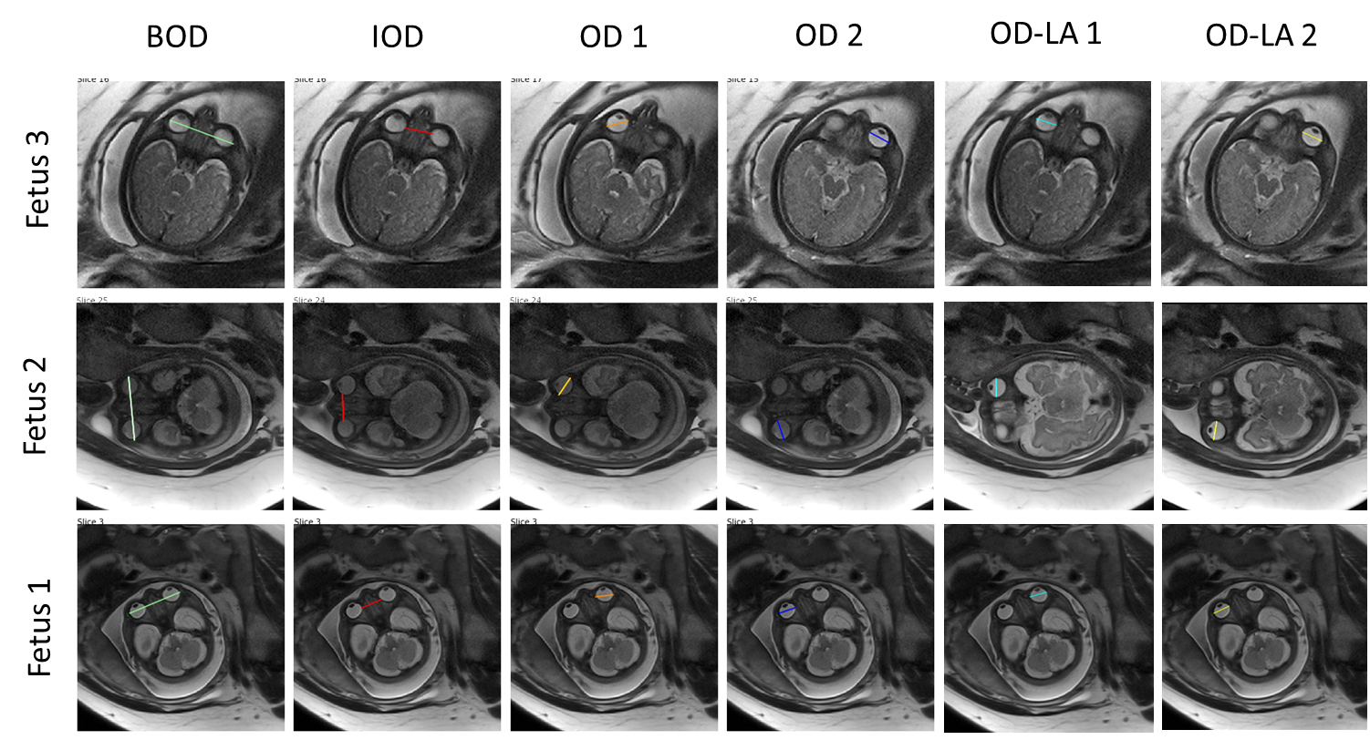
Figure 3: Representative examples of
automatic ocular fetal 2D measurements for three fetuses. OD measurements are
presented for both eyes (OD 1 and 2). Note that the algorithm may choose a different slice for each measurement (for example, IOD, OD1 and OD2 of fetus
3).