Alexey Samsonov1 and Aaron S. Field1
1Radiology, University of Wisconsin-Madison, Madison, WI, United States
1Radiology, University of Wisconsin-Madison, Madison, WI, United States
Improved two-pool MT modeling with calibrated bound proton pool relaxation constraint may allow more specific assessment of macromolecular and paramagnetic tissue components.
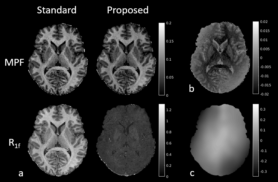
Figure 2. Quantitative
maps estimated using MT modeling with standard and proposed R1b in a healthy volunteer. (a) MPF and R1f. As predicted by simulations (Fig. 1b), standard R1f is dominated by
macromolecular content and therefore resembles MPF. The proposed R1f is much more uniform, likely due
to removing the effects of MPF. Note slightly elevated values in the basal
ganglia, likely due to iron accumulation. (b)
Error between the proposed and standard MPF. Note its high variability with macromolecular
content and B1 field (c), also consistent
with simulations (Fig. 1a).
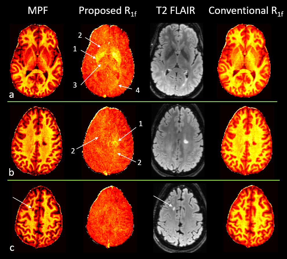
Figure 4. Quantitative
mapping in an MS subject. (a) Proposed
R1f is elevated in deep GM. The
increase is consistent with the known distribution of iron in these regions,
including areas with high (globus pallidus, #1), medium (putamen, #2), and low
(thalamus, #3) iron content. Note elevated R1f
in areas affected by diffuse lesional changes (#4) (as seen on T2-FLAIR and MPF).
(b) The R1f is increased on the rim of the heavily demyelinated lesion (as
revealed by MPF) (#1) and in WM areas affected by more diffuse disease (#2). (c) Example of a lesion without noticeable
increase in R1f.