Gilbert Hangel1, Philipp Lazen2, Matthias Tomschik1, Jonathan Wais1, Eva Hečková2, Lukas Hingerl2, Stephan Gruber2, Bernhard Strasser2, Gregor Kasprian3, Daniela Prayer3, Julia Furtner3, Christoph Baumgartner4, Johannes Koren4, Robert Diehm5, Martha Feucht5, Christian Dorfer1, Ekaterina Pataraia6, Wolfgang Bogner2, Siegfried Trattnig2,7, and Karl Rössler1
1Department of Neurosurgery, Medical University of Vienna, Vienna, Austria, 2High Field MR Centre, Department of Biomedical Imaging and Image-guided Therapy, Medical University of Vienna, Vienna, Austria, 3Division of Neuroradiology and Musculoskeletal Radiology, Department of Biomedical Imaging and Image-guided Therapy, Medical University of Vienna, Vienna, Austria, 4Department of Neurology, Clinic Hietzing, Vienna, Austria, 5Department of Paediatrics and Adolescent Medicine, Medical University of Vienna, Vienna, Austria, 6Department of Neurology, Medical University of Vienna, Vienna, Austria, 7Christian Doppler Laboratory for Clinical Molecular MR Imaging, Vienna, Austria
1Department of Neurosurgery, Medical University of Vienna, Vienna, Austria, 2High Field MR Centre, Department of Biomedical Imaging and Image-guided Therapy, Medical University of Vienna, Vienna, Austria, 3Division of Neuroradiology and Musculoskeletal Radiology, Department of Biomedical Imaging and Image-guided Therapy, Medical University of Vienna, Vienna, Austria, 4Department of Neurology, Clinic Hietzing, Vienna, Austria, 5Department of Paediatrics and Adolescent Medicine, Medical University of Vienna, Vienna, Austria, 6Department of Neurology, Medical University of Vienna, Vienna, Austria, 7Christian Doppler Laboratory for Clinical Molecular MR Imaging, Vienna, Austria
We show the feasibility of CRT-FID-MRSI at 7T for high resolution
metabolic imaging in
epilepsy. We acquired a more extensive metabolic profile at higher resolution
than previous MRSI, with tCho and tCr/NAA/mIns/Glu as most promising markers.
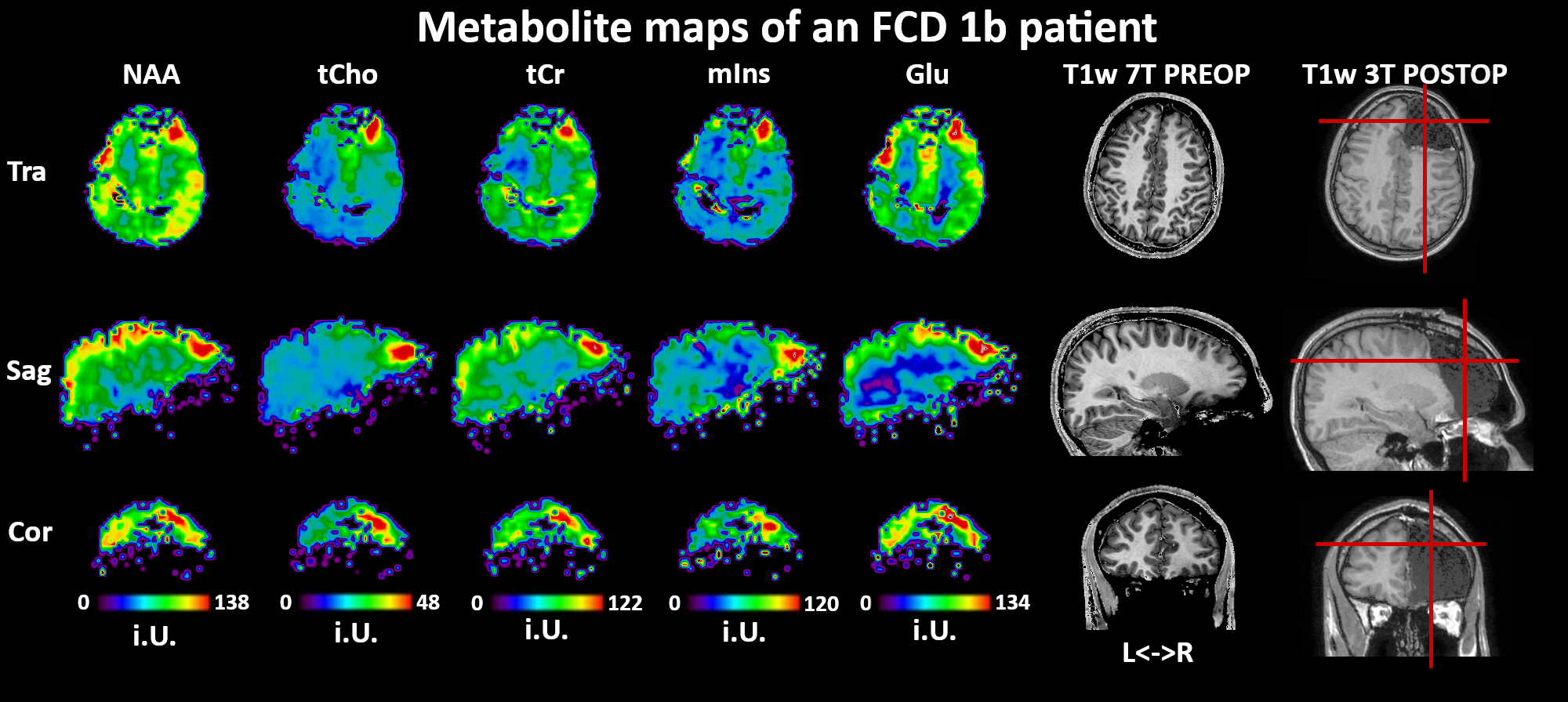
Figure
3: Overview of the possible 3D lesion localisation for multiple metabolites in
patient #2, an FCD 1b patient. The hotspot location corresponds well to later
focal resection.
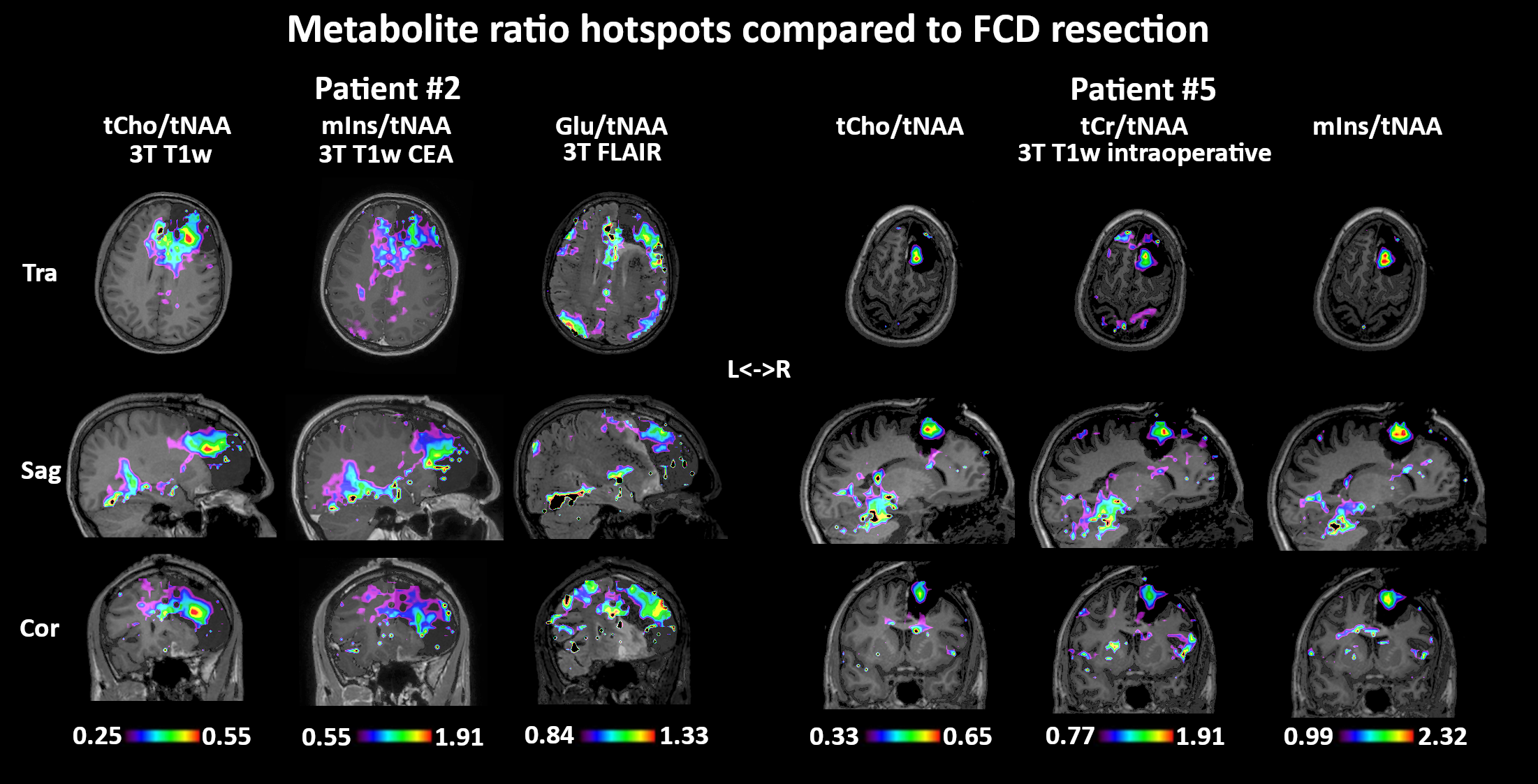
Figure
5: Metabolic ratio maps overlayed to post-resection clinical imaging shows good
correspondence of 7T metabolic imaging to surgical FCD resection.