Hélène Lajous1,2, Tom Hilbert1,3,4, Christopher W. Roy1, Sébastien Tourbier1, Priscille de Dumast1,2, Yasser Alemán-Gómez1, Thomas Yu4, Patric Hagmann1, Mériam Koob1, Vincent Dunet1, Tobias Kober1,3,4, Matthias Stuber1,2, and Meritxell Bach Cuadra1,2,4
1Department of Radiology, Lausanne University Hospital and University of Lausanne, Lausanne, Switzerland, 2CIBM Center for Biomedical Imaging, Lausanne, Switzerland, 3Advanced Clinical Imaging Technology (ACIT), Siemens Healthcare, Lausanne, Switzerland, 4Signal Processing Laboratory 5 (LTS5), Ecole Polytechnique Fédérale de Lausanne (EPFL), Lausanne, Switzerland
1Department of Radiology, Lausanne University Hospital and University of Lausanne, Lausanne, Switzerland, 2CIBM Center for Biomedical Imaging, Lausanne, Switzerland, 3Advanced Clinical Imaging Technology (ACIT), Siemens Healthcare, Lausanne, Switzerland, 4Signal Processing Laboratory 5 (LTS5), Ecole Polytechnique Fédérale de Lausanne (EPFL), Lausanne, Switzerland
We
implemented a flexible numerical framework that simulates realistic clinical magnetic resonance acquisitions of the fetal brain throughout development. We evaluated the robustness of a super-resolution
reconstruction algorithm to noise and motion in the simulated fetal brain images.
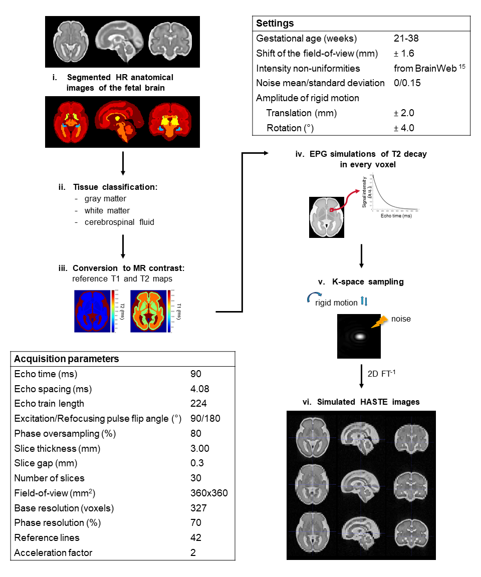
Simulation pipeline of HASTE acquisitions from segmented high-resolution
anatomical images of the fetal brain. This framework offers a great flexibility
in the choice of the sequence parameters but also other settings such as the
age of the fetus, the SNR of the acquisitions, and the
amplitude of fetal motion.
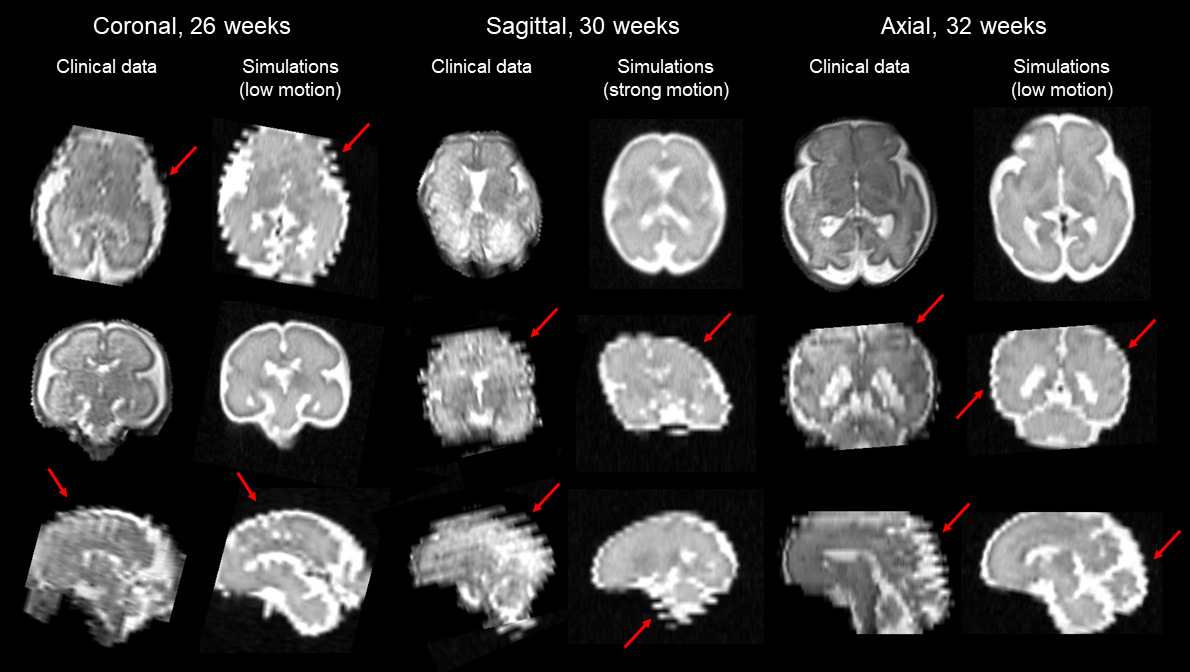
Visual inspection and comparison between simulated HASTE images and
clinical acquisitions at three different gestational ages (26, 30 and 32
weeks). Various levels of fetal movement can be observed. Arrows point out typical out-of-plane motion patterns.