Yoshiharu Ohno1,2,3, Masao Yui4, Takeshi Yoshikawa3,5, Daisuke Takenaka5, Kaori Yamamoto4, Yoshimori Kassai4, Kazuhiro Murayama2, and Hiroshi Toyama1
1Radiology, Fujita Health University School of Medicine, Toyoake, Japan, 2Joint Research Laboratory of Advanced Medical Imaging, Fujita Health University School of Medicine, Toyoake, Japan, 3Division of Functional and Diagnostic Imaging Research, Department of Radiology, Kobe University Graduate School of Medicine, Kobe, Japan, 4Canon Medical Systems Corporation, Otawara, Japan, 5Diagnostic Radiology, Hyogo Cancer Center, Akashi, Japan
1Radiology, Fujita Health University School of Medicine, Toyoake, Japan, 2Joint Research Laboratory of Advanced Medical Imaging, Fujita Health University School of Medicine, Toyoake, Japan, 3Division of Functional and Diagnostic Imaging Research, Department of Radiology, Kobe University Graduate School of Medicine, Kobe, Japan, 4Canon Medical Systems Corporation, Otawara, Japan, 5Diagnostic Radiology, Hyogo Cancer Center, Akashi, Japan
Pulmonary MR imaging with UTE has
a potential to be applied for lung cancer screening as well as CT.
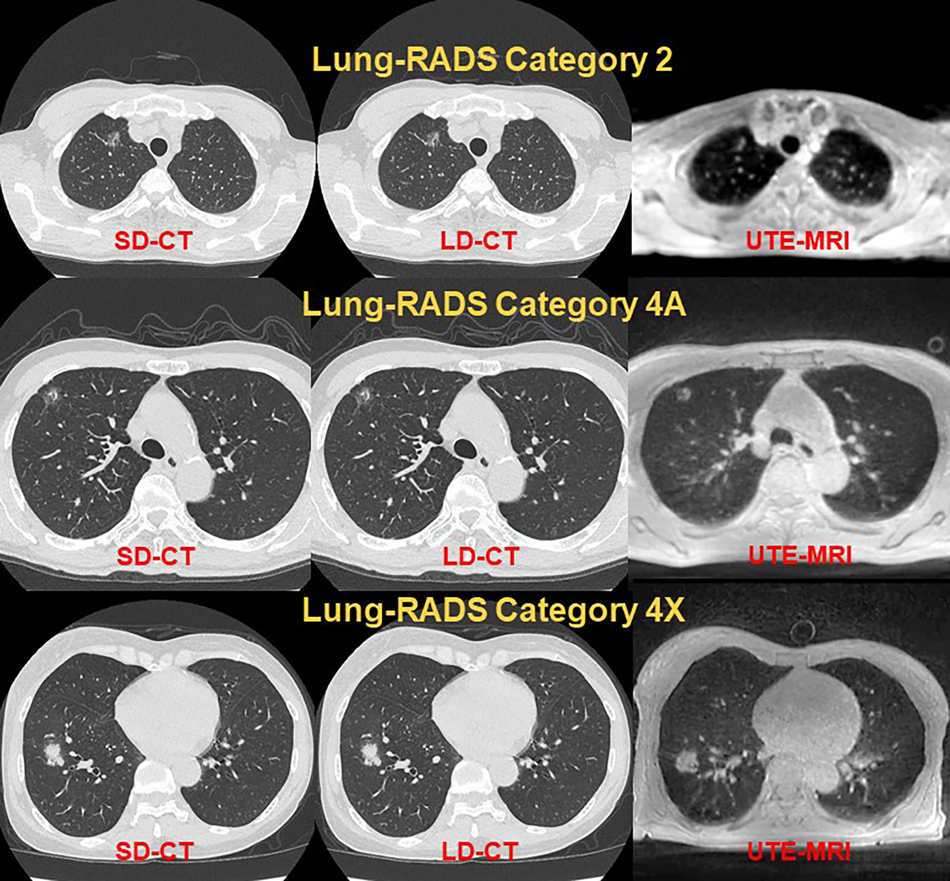
Figure 1. Examples for standard-dose (SD-CT: Left), low-dose CT (LD-CT: Middle) and pulmonary MR imaging with UTE (UTE-MRI: Right) in Lung-RADS Category 2 (1st line), catefory 4A (2nd line) and Category 4X (3rd line).
Each method has no difference of radiological findings as well as Lung-RADS classification in each subject.
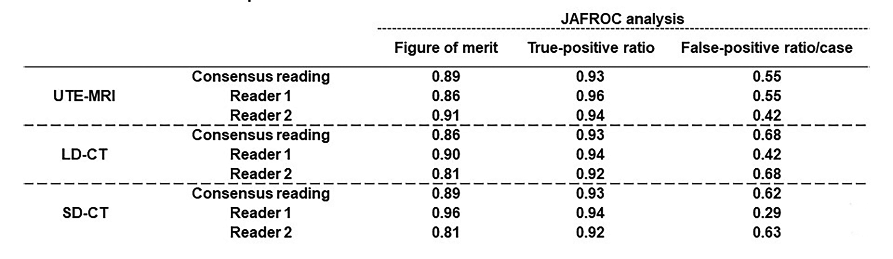
Figure 2. Results of JAFROC analysis for lung nodule detection.
There were no significant differences of figure of merit, true-positive ratio and false-positive ratio/case among all methods.