Lincoln Craven-Brightman1, Thomas O'Reilly2, Benjamin Menkuec3, Marcus Prier4, Rubén Pellicer-Guridi5,6, Joseba Alonso5,6, Lawrence L. Wald1,7, Maxim Zaitsev8, Jason Stockmann1,7, Thomas Witzel9, Andrew Webb2, and Vlad Negnevitsky10
1A.A. Martinos Center for Biomedical Imaging, Department of Radiology, Massachusetts General Hospital, Charlestown, MA, United States, 2Department of Radiology, Leiden University Medical Center, Leiden, Netherlands, 3University of Applied Sciences and Arts Dortmund, Dortmund, Germany, 4Otto von Guericke Universität Magdeburg, Magdeburg, Germany, 5Universitat Politècnica de València, Valencia, Spain, 6Spanish National Research Council, Valencia, Spain, 7Harvard Medical School, Boston, MA, United States, 8Medizinische Universität Wien, Vienna, Austria, 9Qbio Inc, San Carlos, CA, United States, 10Independent researcher, Zürich, Switzerland
1A.A. Martinos Center for Biomedical Imaging, Department of Radiology, Massachusetts General Hospital, Charlestown, MA, United States, 2Department of Radiology, Leiden University Medical Center, Leiden, Netherlands, 3University of Applied Sciences and Arts Dortmund, Dortmund, Germany, 4Otto von Guericke Universität Magdeburg, Magdeburg, Germany, 5Universitat Politècnica de València, Valencia, Spain, 6Spanish National Research Council, Valencia, Spain, 7Harvard Medical School, Boston, MA, United States, 8Medizinische Universität Wien, Vienna, Austria, 9Qbio Inc, San Carlos, CA, United States, 10Independent researcher, Zürich, Switzerland
A low cost programmable MR console, iterating on the Open Source Console for Realtime Acquisition (OCRA) characterized and tested in various setups at multiple sites for both educational and research applications. Notable points are a PulSeq interpreter and the first low-field in-vivo scans.
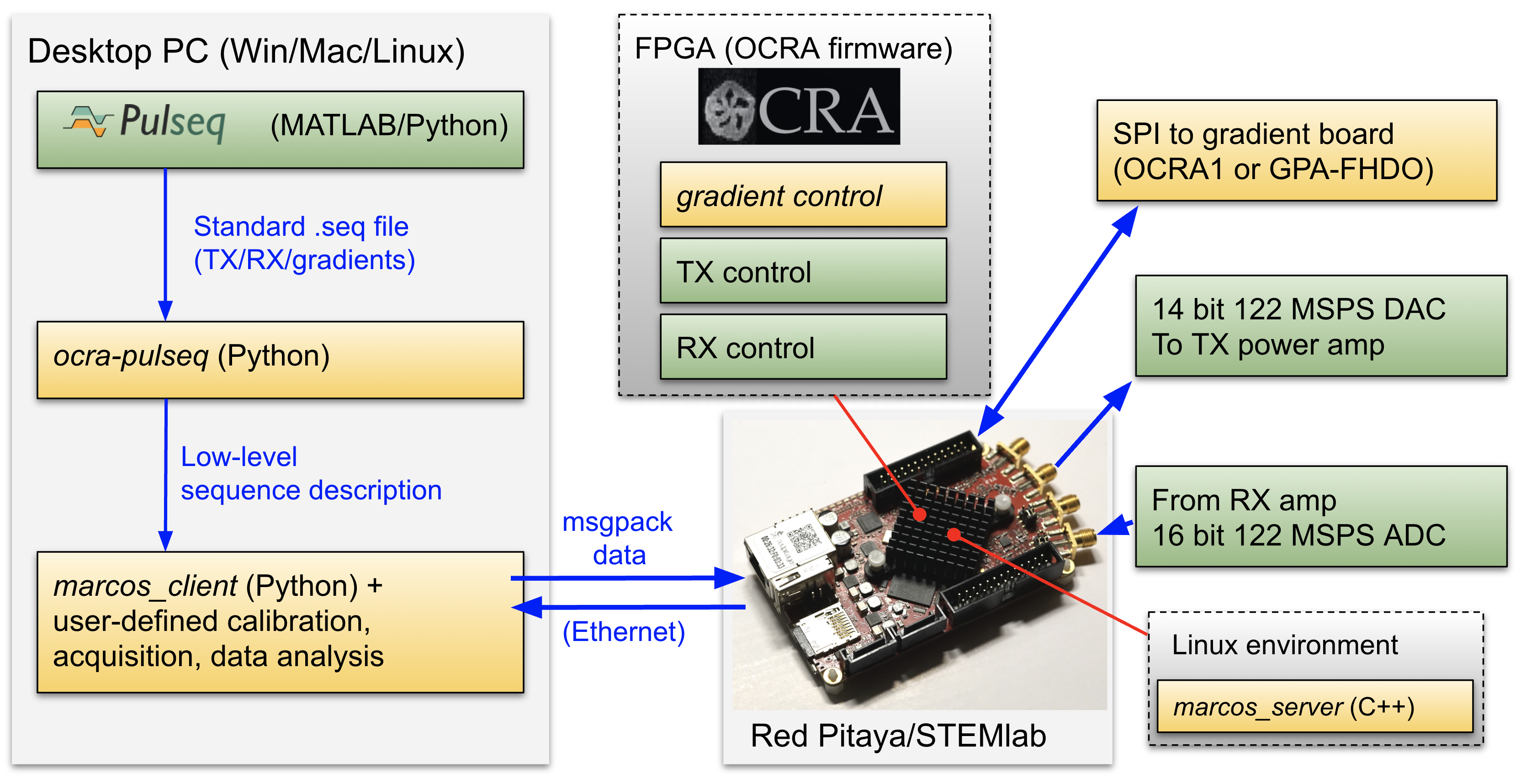
Figure 1. Software and hardware stack of our OCRA package. A standard PulSeq description (.seq) file is converted by the ocra-pulseq and marcos_client libraries into data that can be executed on the open-source OCRA console. Yellow boxes show the novel elements in the stack: ocra-pulseq and marcos_client Python libraries, the new server and gradient firmware in the OCRA system, and the new OCRA1 DAC and GPA-FHDO gradient boards.
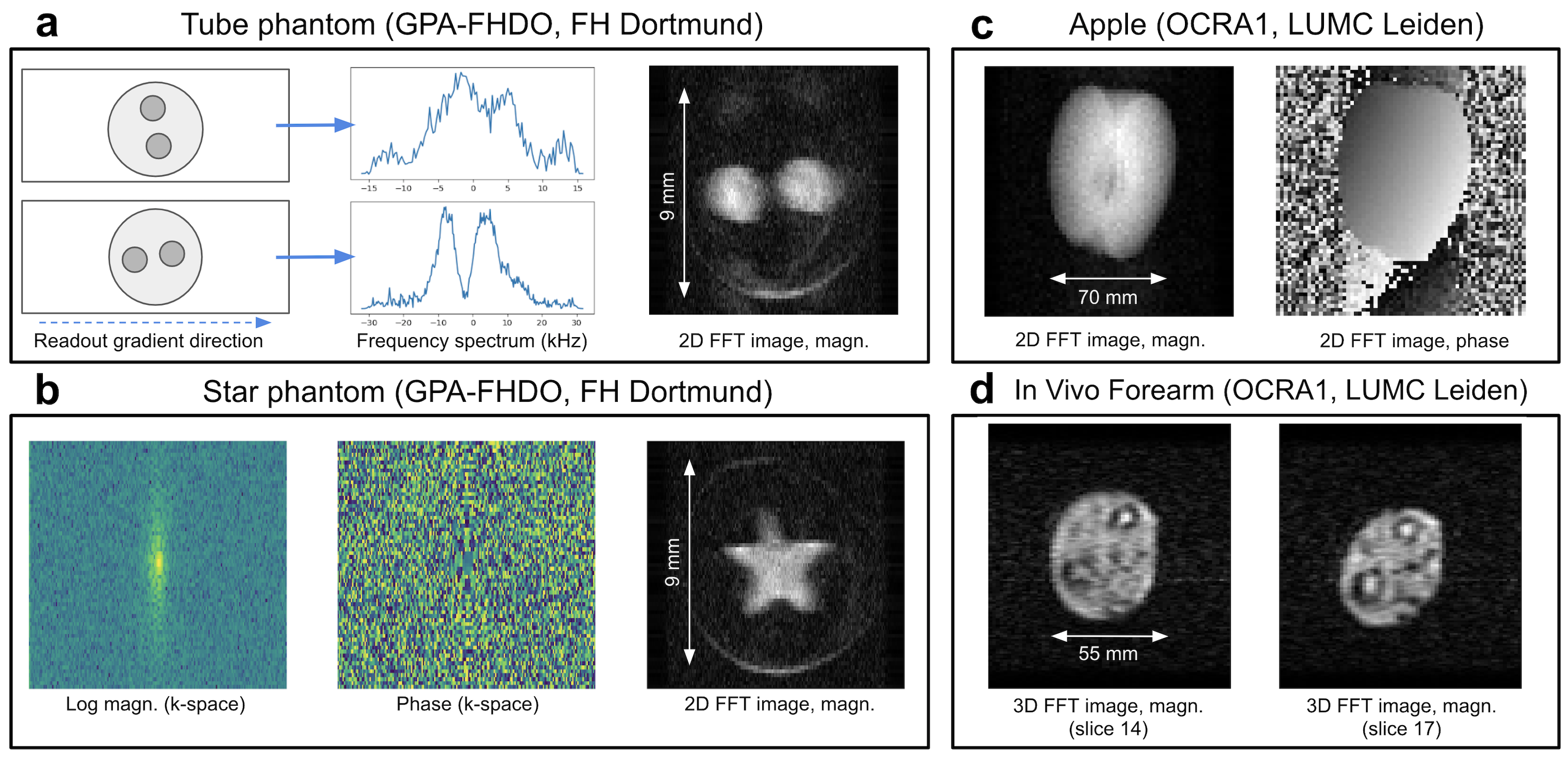
Figure 4. Example imaging data acquired with OCRA console. (a) 1D projection imaging of a 1-cm FOV two-tube phantom for use in MRI educational curriculum on a tabletop scanner, along with 2D FFT reconstruction. (b) K-space data and a 2D image of a star phantom are also shown. (c) Phase and magnitude of a 2D turbo spin echo acquisition of an apple, and (d) two slides of a 3D turbo spin echo acquisition of a forearm in vivo, both acquired on the Leiden brain scanner platform.