Vikas Chauhan1, Sandeep Kaushik2, Florian Wiesinger2, Cristina Cozzini2, Michael Carl2, Maggie Fung2, Bhairav Mehta2, Bejoy Thomas1, and Kesavadas Chandrasekharan1
1Imaging Sciences and Interventional Radiology, Sree Chitra Tirunal Institute for Medical Sciences and Technology, Thiruvananthapuram, India, 2GE Healthcare, GE Healthcare, Bangalore, India
1Imaging Sciences and Interventional Radiology, Sree Chitra Tirunal Institute for Medical Sciences and Technology, Thiruvananthapuram, India, 2GE Healthcare, GE Healthcare, Bangalore, India
Zero Time of Echo imaging technique is able to image the bone and its image quality is comparable to the real CT scan images. The advantage of this is that during the same MRI bone imaging can be also done and patient need not go for another investigation that involve ionising radiation.
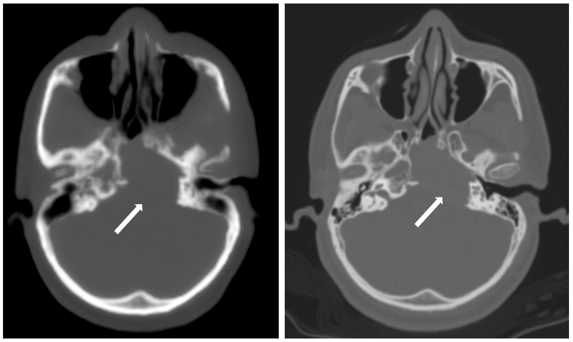
Figure 1: Left petrous Giant cell tumor. PseudoCT image (L) clearly depicting the lesion and its margins as compared with CT image (R).
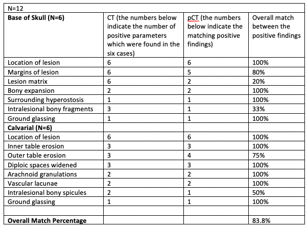
Table 1: Results of the image analysis between PseudoCT and CT scan images.