Yida Wang1, YinQiao Yi1, Haijie Wang1, Changan Chen2, Yingfang Wang2, Guofu Zhang2, He Zhang2, and Guang Yang1
1East China Normal University, Shanghai Key Laboratory of Magnetic Resonance, Shanghai, China, 2Department of Radiology, Obstetrics and Gynecology Hospital, Fudan University, Shanghai, China
1East China Normal University, Shanghai Key Laboratory of Magnetic Resonance, Shanghai, China, 2Department of Radiology, Obstetrics and Gynecology Hospital, Fudan University, Shanghai, China
We proposed a deep
learning (DL) approach to segment ovarian lesion and differentiate ovarian malignant
from borderline tumors in MR Imaging. The trained DL network model could help
to identify and categorize ovarian masses with a high accuracy.
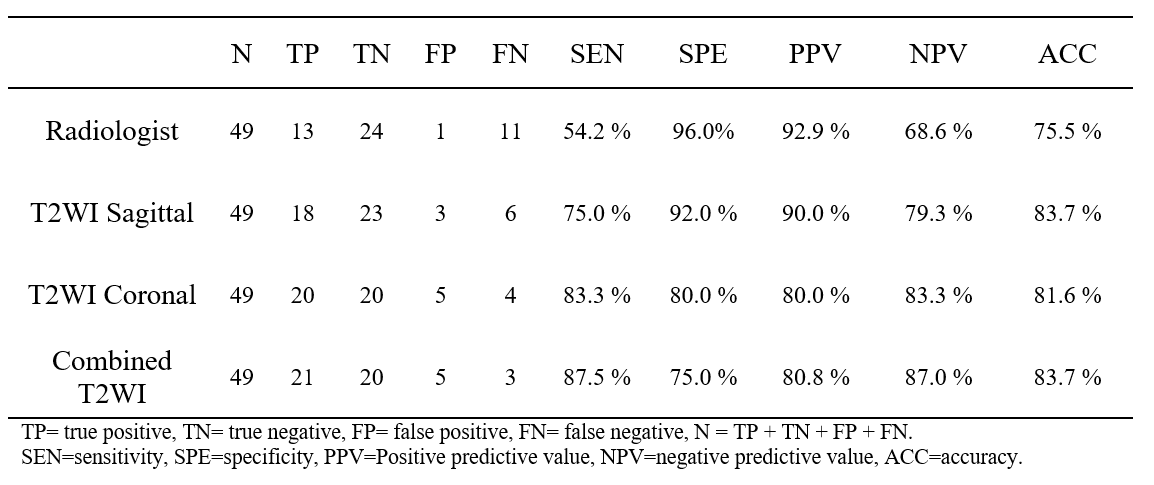
Table 1. Comparison of diagnostic
performance of the radiologist and CNN models in ovarian mass discrimination based
on MR images in the testing group.
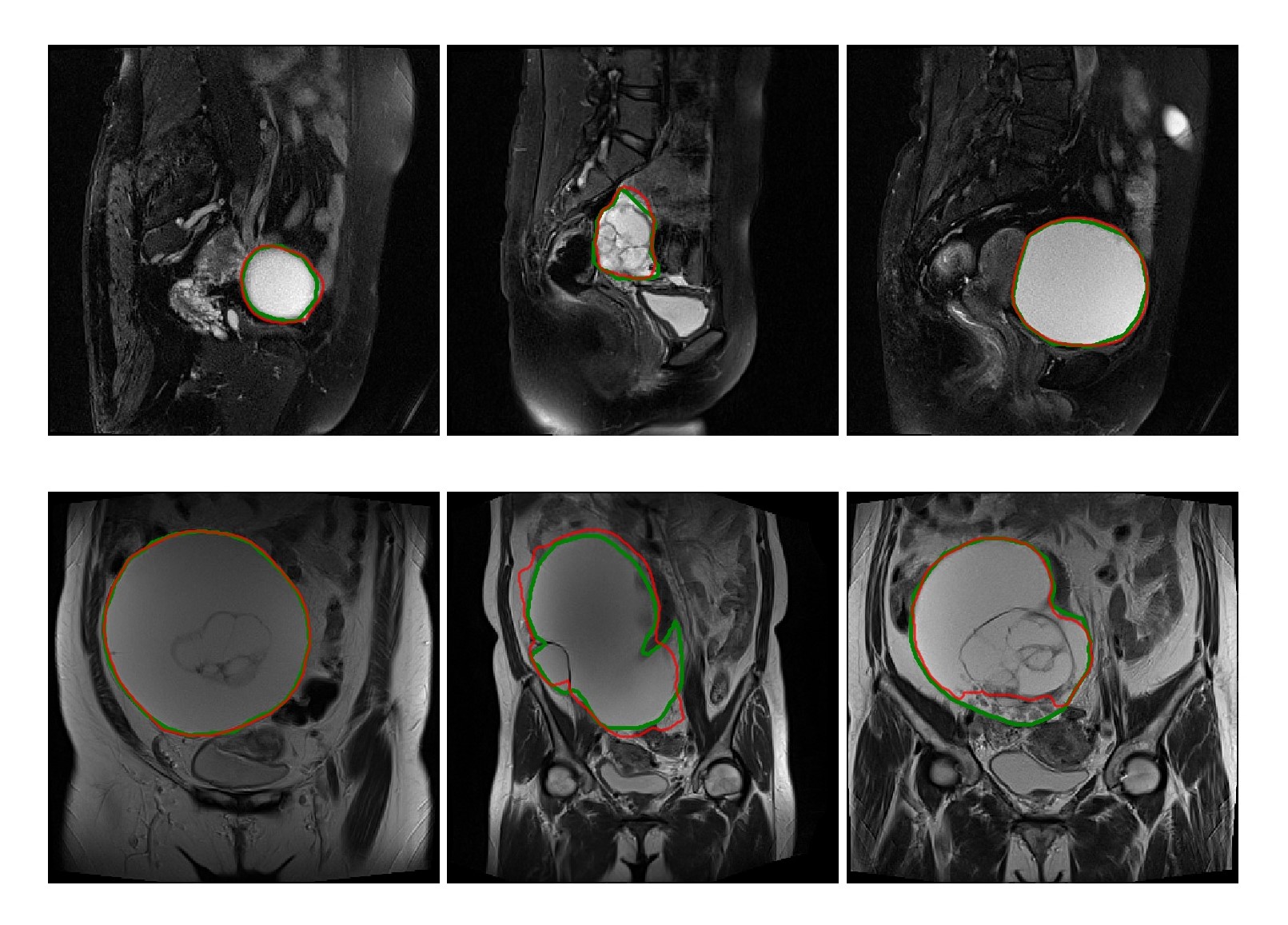
Figure 3. The
segmentation results on MR images. The segmented ovarian tumor regions by
U-net++ network (the red line) and radiologist (the green line, ground truth)
are shown on sagittal T2WI (upper row) and T2WI coronal (down
row) MR images.