Shubhangi Agarwal1, Jinny Sun1, Emilie Decavel-Bueff1, Robert A Bok1, Romelyn Delos Santos1, Mark Van Criekinge1, Rahul Aggarwal2, Daniel B Vigneron1, Donna Peehl1, John Kurhanewicz1, and Renuka Sriram1
1Department of Radiology and Biomedical Imaging, University of California, San Francisco, San Francisco, CA, United States, 22Division of Hematology/Oncology, Department of Medicine, University of California, San Francisco, San Francisco, CA, United States
1Department of Radiology and Biomedical Imaging, University of California, San Francisco, San Francisco, CA, United States, 22Division of Hematology/Oncology, Department of Medicine, University of California, San Francisco, San Francisco, CA, United States
Hyperpolarized [1-13C] pyruvate conversion to lactate may be used to indicate early response to carboplatin in small cell neuroendocrine prostate cancer models in the bone.
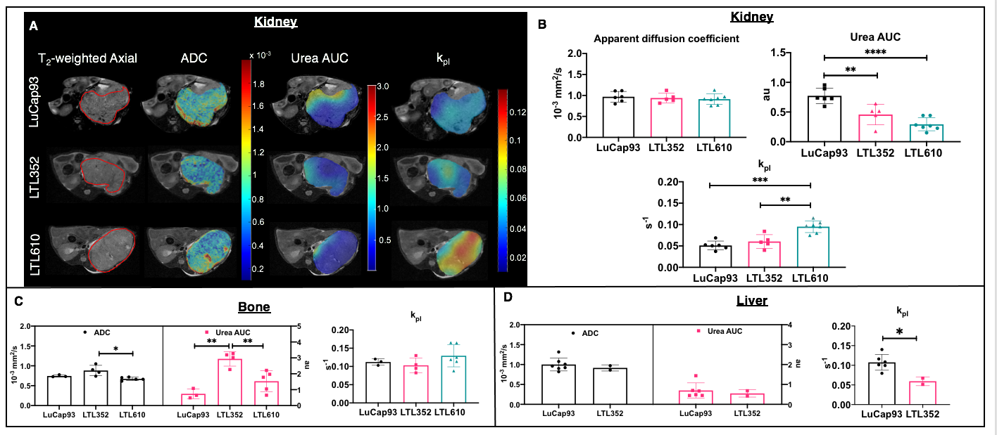
Figure 1: Representative T2-weighted images of PDXs implanted in kidney overlaid with ADC, urea AUC and kpl maps (A). The tumor is delineated with red line. Mean apparent diffusion coefficients, urea AUCs and kpl of kidney (B), bone (C) and liver tumors (D). (Note: significance shown as p values. * p<0.05, **p<0.01 and ***p<0.001)
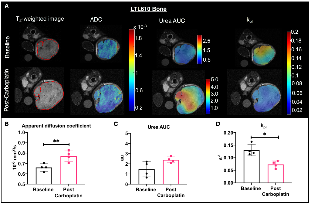
Figure 3: Representative T2-weighted images of LTL610 PDXs implanted in bone at baseline and post-carboplatin overlaid with ADC, urea AUC and kpl maps (A). Mean apparent diffusion coefficients (B), urea AUC (C) and kpl (D) at baseline and post-carboplatin. (Note: significance shown as p values. * p<0.05 and **p<0.01.)