Joachim Snellings1, Kader Avan1, Marcus Markowski2, Bernd Hamm1, Patrick Asbach1, Carsten Warmuth1, Mehrgan Shahryari1, Heiko Tzschätzsch1, Ingolf Sack1, and Jürgen Braun3
1Institute of Radiology, Charité Üniversitätsmedizin, Berlin, Germany, 2School of Medicine & Klinikum Rechts der Isar, Technical University of Munich, Munich (TUM), München, Germany, 3Institut für Medizinische Informatik, Charité Üniversitätsmedizin, Berlin, Germany
1Institute of Radiology, Charité Üniversitätsmedizin, Berlin, Germany, 2School of Medicine & Klinikum Rechts der Isar, Technical University of Munich, Munich (TUM), München, Germany, 3Institut für Medizinische Informatik, Charité Üniversitätsmedizin, Berlin, Germany
Clinical MR scanners with
modern MRE hardware and ultrafast MRE sequences allow the determination of
viscoelastic parameters of implanted tumors in murine models.

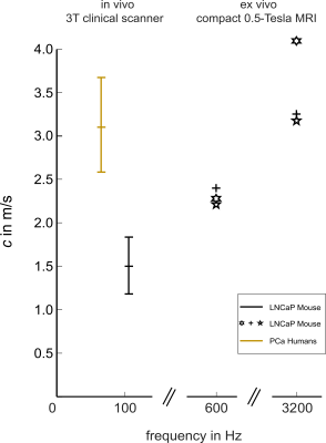
Figure 5: Overview of measured SWS (c) in patients and in the LNCap murine model. Comparison of
the k-MDEV-based group mean SWS (±Std) determined in vivo on clinical scanners,
show that PCa’s in their native environment(4) are stiffer (3.1±0.6m/s, frequency-range 60-80 Hz) than the implanted LNCap tumors in the murine-model (1.5±0.3 m/s, frequency-range of 80-130Hz).Considering the
frequency dispersion of c in the ex-vivo study, a good agreement can be
found with in-vivo data. See the Result section for detailed SWS values measured ex-vivo.

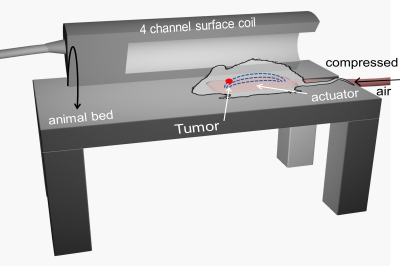
Figure 1: Experimental
setup. To induce shear waves the anesthetized animals
are positioned directly on a passive pressurized air driven actuator (size:80×40×10mm3).
The acquisition coil, here sketched as a halved hollow cylinder (inner diameter 47mm), is positioned over the animal. The table height is adjusted so
that the animals can be positioned in the isocenter of the magnet.
