Wilburn E Reddick1, Jared J Sullivan1, John O Glass1, Yian Guo2, Julie H Harreld1, Yimei Li2, Giles W Robinson3, Amar Gajjar3, and Thomas E Merchant4
1Department of Diagnostic Imaging, St. Jude Children's Research Hospital, Memphis, TN, United States, 2Department of Biostatistics, St. Jude Children's Research Hospital, Memphis, TN, United States, 3Department of Oncology, St. Jude Children's Research Hospital, Memphis, TN, United States, 4Department of Radiation Oncology, St. Jude Children's Research Hospital, Memphis, TN, United States
1Department of Diagnostic Imaging, St. Jude Children's Research Hospital, Memphis, TN, United States, 2Department of Biostatistics, St. Jude Children's Research Hospital, Memphis, TN, United States, 3Department of Oncology, St. Jude Children's Research Hospital, Memphis, TN, United States, 4Department of Radiation Oncology, St. Jude Children's Research Hospital, Memphis, TN, United States
FA measures at baseline demonstrated an immediate decline due to tumor
and surgery, which is then accentuated by
irradiation followed by a partial recovery, which was attenuated in
patients receiving higher doses.
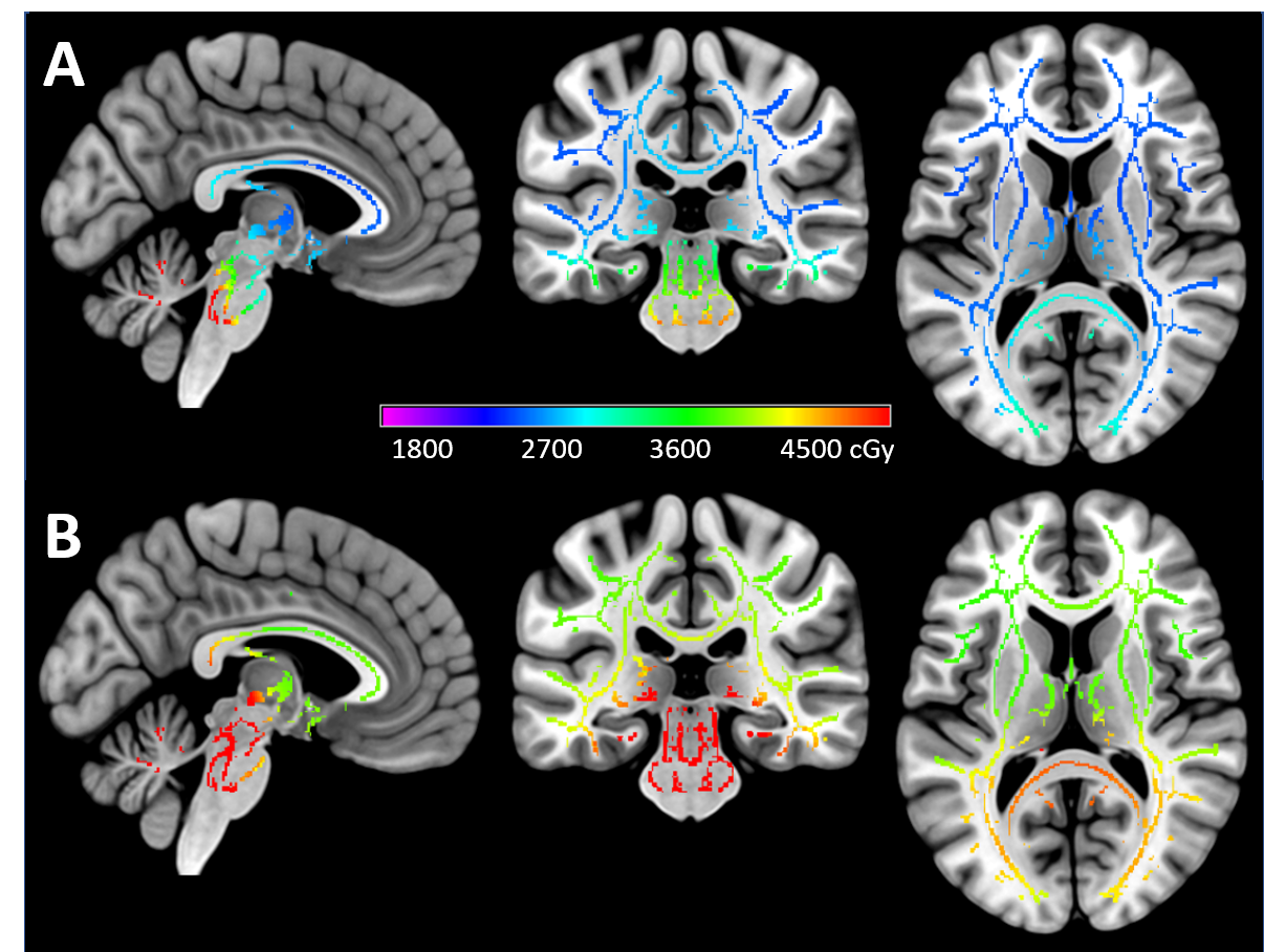
Figure 1: Representative individual dosimetry mapped in cGy onto the TBSS skeleton
for an average-risk (A) and high-risk (B) patient.
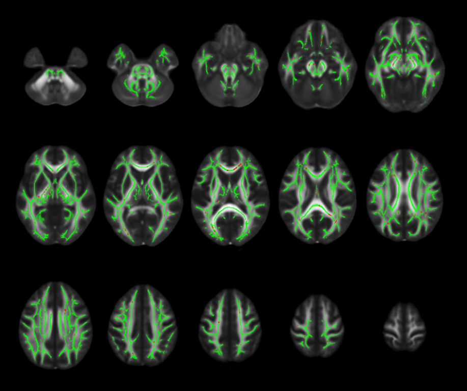
Figure 5: TBSS results for interaction of dose with longitudinal
evolution of the FA after irradiation. The skeleton is shown green overlaid on
the average FA with red voxels demarcating significant negative interaction
between dose and the positive change over time (slope) indicating that higher
doses attenuated the rate of recovery. Distributions of these voxels were
primarily in the cerebral peduncles, internal capsule, posterior thalamic
radiation, corpus callosum, and corona radiata including the superior longitudinal
fasciculus.