Samuel Bobholz1, Allison Lowman2, Michael Brehler2, Savannah Duenweg1, Fitzgerald Kyereme2, Elizabeth Cochran3, Jennifer Connelly4, Wade Mueller5, Mohit Agarwal2, Darren O'Neill2, Anjishnu Banerjee6, and Peter LaViolette2,7
1Biophysics, Medical College of Wisconsin, Milwaukee, WI, United States, 2Radiology, Medical College of Wisconsin, Milwaukee, WI, United States, 3Pathology, Medical College of Wisconsin, Milwaukee, WI, United States, 4Neurology, Medical College of Wisconsin, Milwaukee, WI, United States, 5Neurosurgery, Medical College of Wisconsin, Milwaukee, WI, United States, 6Biostatistics, Medical College of Wisconsin, Milwaukee, WI, United States, 7Biomedical Engineering, Medical College of Wisconsin, Milwaukee, WI, United States
1Biophysics, Medical College of Wisconsin, Milwaukee, WI, United States, 2Radiology, Medical College of Wisconsin, Milwaukee, WI, United States, 3Pathology, Medical College of Wisconsin, Milwaukee, WI, United States, 4Neurology, Medical College of Wisconsin, Milwaukee, WI, United States, 5Neurosurgery, Medical College of Wisconsin, Milwaukee, WI, United States, 6Biostatistics, Medical College of Wisconsin, Milwaukee, WI, United States, 7Biomedical Engineering, Medical College of Wisconsin, Milwaukee, WI, United States
MP-MRI
signatures of cellularity, CYT, and ECF capture a modest degree of variance
using autopsy samples as ground truth, with stronger radio-pathomic
relationships observed in non-GBM patients than GBM patients.
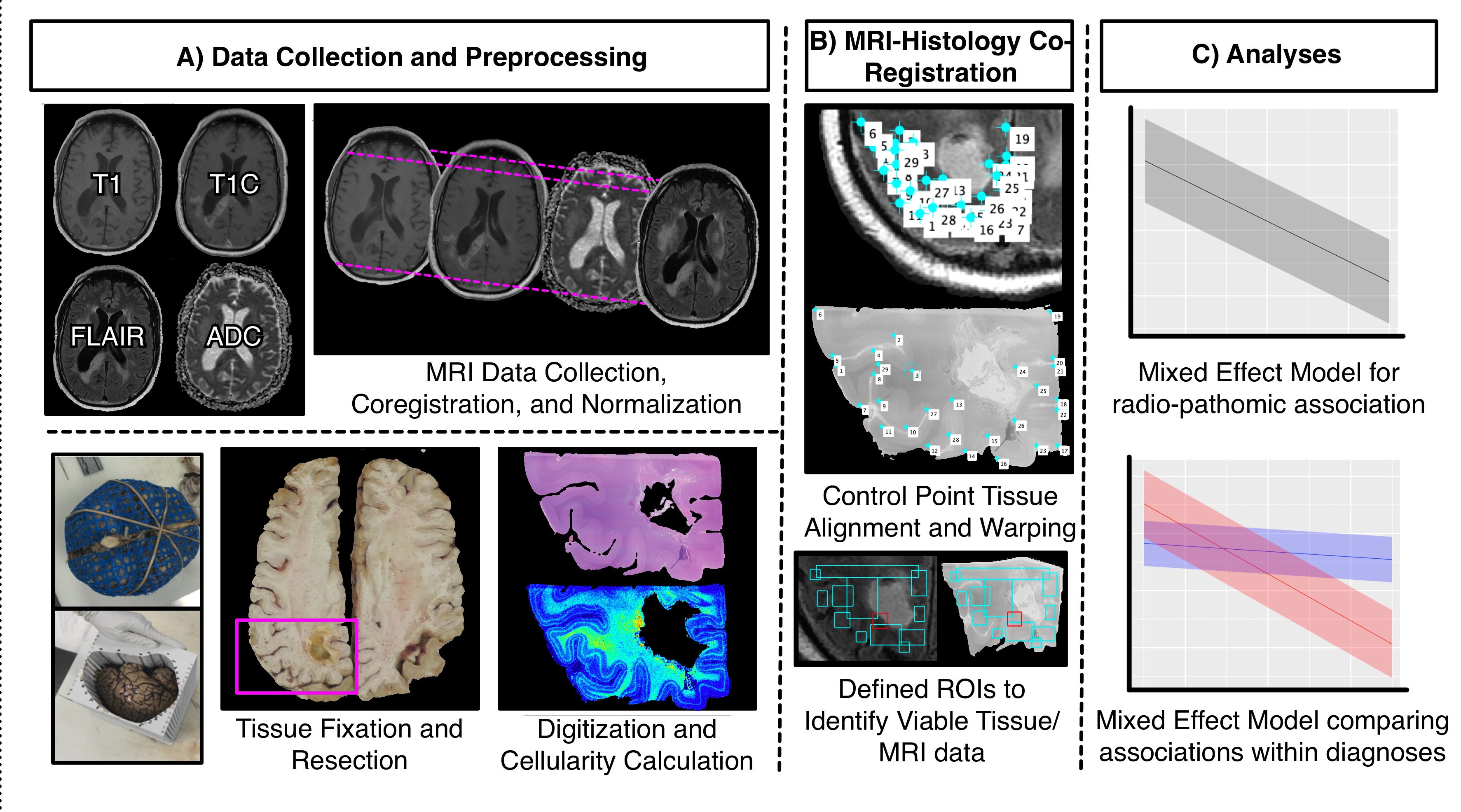
Schematic representation of the study structure including A) data collection for MRI and tissue modalities and associated preprocessing, B) MR-histology coregistration, and C) mixed effect analysis structure
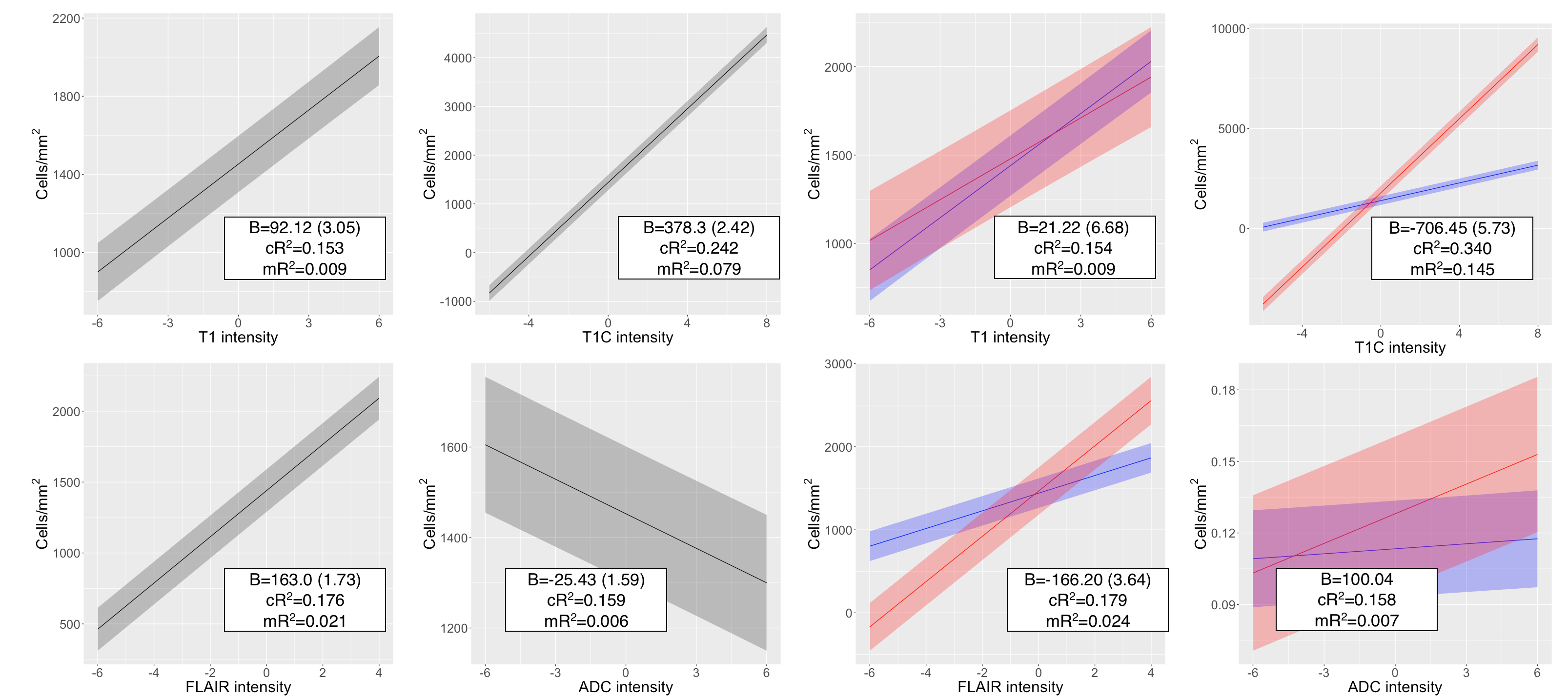
Mixed effect model results for cell density. The left-hand plots represent the first order models, whereas the right hand results represent the interaction between MR intensity and diagnosis, where blue = GBM and red = non-GBM.