Manabu Kinoshita1, Hideyuki Arita1, Masamichi Takahashi2, Takehiro Uda3, Junya Fukai4, Kenichi Ishibashi5, Noriyuki Kijima1, Ryuichi Hirayama1, Mio Sakai6, Atsuko Arisawa7, Hiroto Takahashi7, Katsuyuki Nakanishi6, Naoki Kagawa1, Kouichi Ichimura8, Yonehiro Kanemura9, Yoshitaka Narita2, and Haruhiko Kishima1
1Neurosurgery, Osaka University Graduate School of Medicine, Suita, Japan, 2Neurosurgery and Neuro-Oncology, National Cancer Center Hospital, Tokyo, Japan, 3Neurosurgery, Osaka City University Graduate School of Medicine, Osaka, Japan, 4Neurological Surgery, Wakayama Medical University, Wakayama, Japan, 5Neurosurgery, Osaka City General Hospital, Osaka, Japan, 6Diagnostic Radiology, Osaka International Cancer Institute, Osaka, Japan, 7Radiology, Osaka University Graduate School of Medicine, Suita, Japan, 8Division of Brain Tumor Translational Research, National Cancer Center Research Institute, Tokyo, Japan, 9Biomedical Research and Innovation, National Hospital Organization Osaka National Hospital, Osaka, Japan
1Neurosurgery, Osaka University Graduate School of Medicine, Suita, Japan, 2Neurosurgery and Neuro-Oncology, National Cancer Center Hospital, Tokyo, Japan, 3Neurosurgery, Osaka City University Graduate School of Medicine, Osaka, Japan, 4Neurological Surgery, Wakayama Medical University, Wakayama, Japan, 5Neurosurgery, Osaka City General Hospital, Osaka, Japan, 6Diagnostic Radiology, Osaka International Cancer Institute, Osaka, Japan, 7Radiology, Osaka University Graduate School of Medicine, Suita, Japan, 8Division of Brain Tumor Translational Research, National Cancer Center Research Institute, Tokyo, Japan, 9Biomedical Research and Innovation, National Hospital Organization Osaka National Hospital, Osaka, Japan
This study discovered
that FLAIR acquisition with TI shorter than 2400 ms in 3T could
Improve the
detectability of IDHmt, non-CODEL astrocytomas. Tuning TI for FLAIR acquisition is such a simple
technique that clinicians can easily incorporate into daily workflow of glioma imaging.
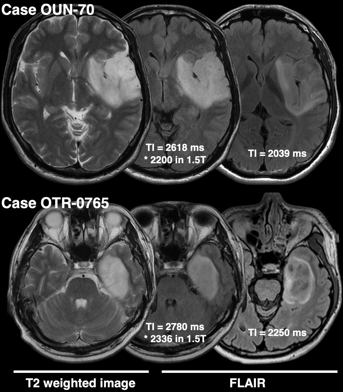
This figure shows two representative IDHmt,
non-CODEL astrocytomas that had two FLAIR images scanned with different TIs.
Both cases highlight the importance of TI for FLAIR acquisition in terms of
visualization of the T2-FLAIR mismatch sign.
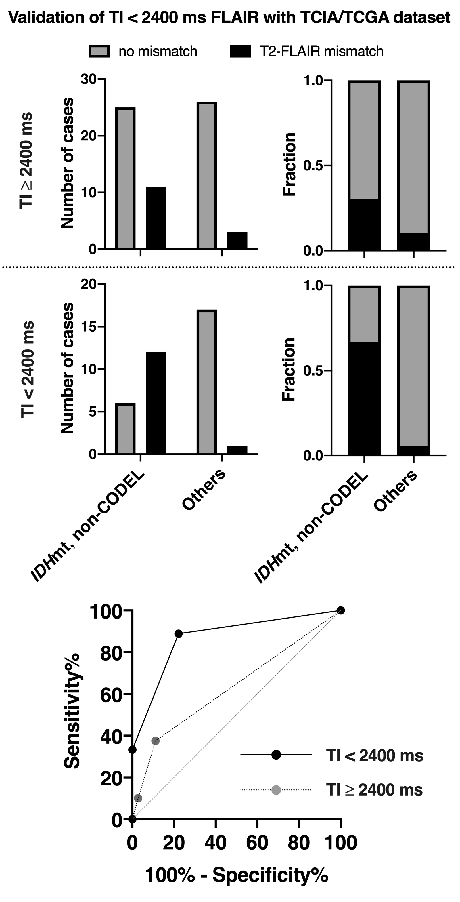
The figures show the frequencies of
presence or absence of the T2-FLAIR mismatch sign found in the TCIA/TCGA LrGG
cohort. We divided the cohort into two groups according to the TI for FLAIR
acquisition. The T2-FLAIR mismatch sign was more frequently positive for or IDHmt,
non-CODEL astrocytomas if we acquired FLAIR with TI shorter than 2400 ms. The
ROC curve for identifying IDHmt, non-CODEL astrocytomas is presented at
the lower panel. The area under the curve improved from 0.63 to 0.87 87 if we
acquired FLAIR with TI shorter than 2400 ms.