Fatemeh Arzanforoosh1, Paula L. Croal2, Karin Van Garderen1, Marion Smits1, Michael A. Chappell2, and Esther A.H. Warnert1
1Department of Radiology & Nuclear Medicine, ErasmusMC, Rotterdam, Netherlands, 2Radiological Sciences, Division of Clinical Neurosciences, University of Nottingham, Nottingham, United Kingdom
1Department of Radiology & Nuclear Medicine, ErasmusMC, Rotterdam, Netherlands, 2Radiological Sciences, Division of Clinical Neurosciences, University of Nottingham, Nottingham, United Kingdom
In summary, this work recommends application of a pre-bolus combined
with BSW leakage correction in enhancing glioma for vessel size estimation,
while eliminating the need for leakage correction for nonenhancing glioma.
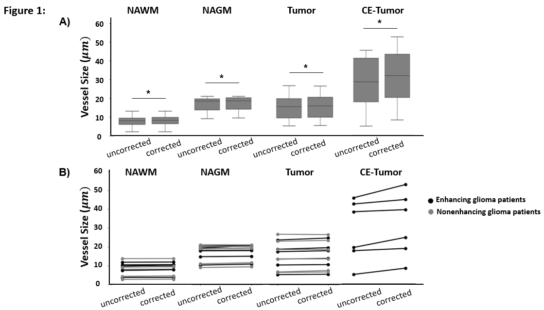
Figure 1: A)
Group averages of corrected and uncorrected vessel size measurements within 4
ROIs: NAWM, NAGM, Tumor,
CE-Tumor.
Note that CE-Tumor
ROI was delineated only for enhancing glioma patients
(6 patients) , B)
Scatter plots of corrected and uncorrected vessel size measurements
,
across subject within 4 ROIs (individual subject’s data are connected by solid
lines). *
represents significant p-value calculated from Student’s t-test (p
< 0.05)
.
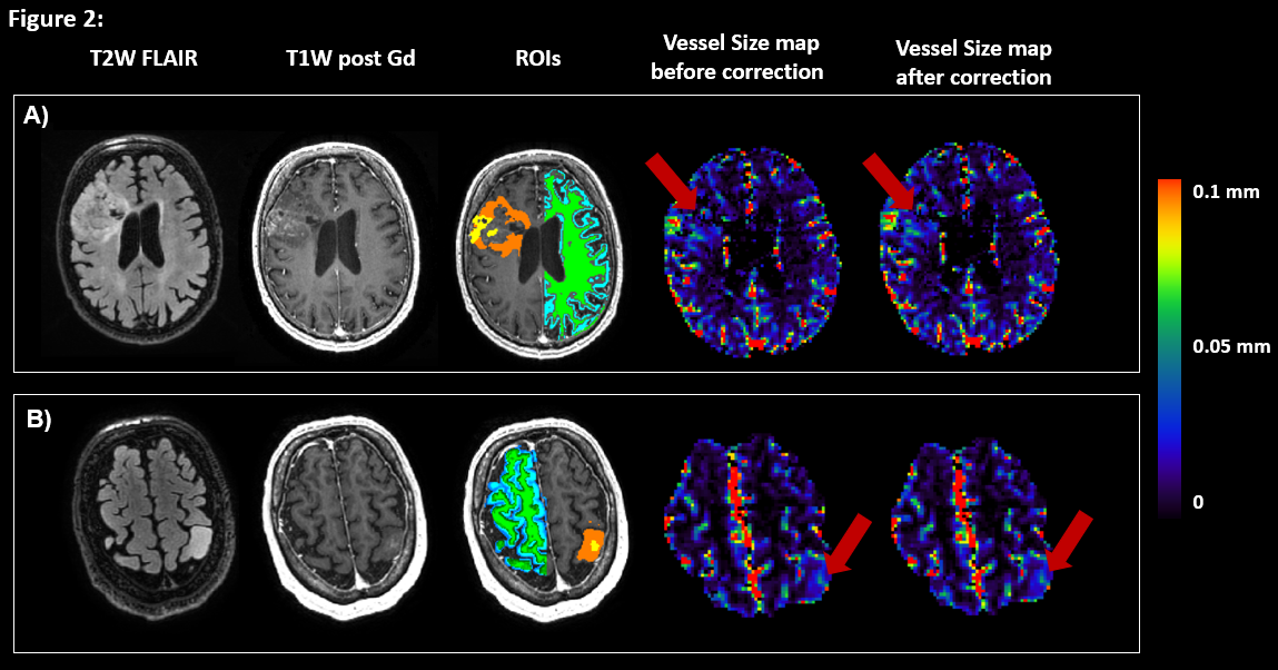
Figure 2: Single slice of exemplary MRI images
of two patients
with enhancing gliomas ; A) Patient 01 and
B) Patient 03. The images from left to right respectively are : T2W FLAIR; post
contrast T1W; post contrast T1W with regions of interest, ROIs, overlaid (contrast-enhanced
tumor (CE-Tumor) in yellow, tumor in orange,
normal-appearing white matter (NAWM)
in green,
and normal-appearing grey matter (NAGM) in blue), calculated vessel
size
map (before application of leakage correction) and calculated
vessel size
map (after application of leakage correction).