Nan Zhang1, Qingwei Song2, Ailian Liu2, Haonan Zhang2, Renwang Pu2, Jiazheng Wang3, and Zhiwei Shen3
1The First Affilliated Hospital of Dalian Medical University, Dalian, China, 2The First Affiliated Hospital of Dalian Medical University, Dalian, China, 3Philips Healthcare, Beijing, China, Beijing, China
1The First Affilliated Hospital of Dalian Medical University, Dalian, China, 2The First Affiliated Hospital of Dalian Medical University, Dalian, China, 3Philips Healthcare, Beijing, China, Beijing, China
The study aims to explore the feasibility of compressed
SENSE with different acceleration factors in 3D brain APTw imaging. Based on
the results, an accelerator factor of 5 is recommended with acceptable image
quality and significantly reduced scan (less than 2? min).
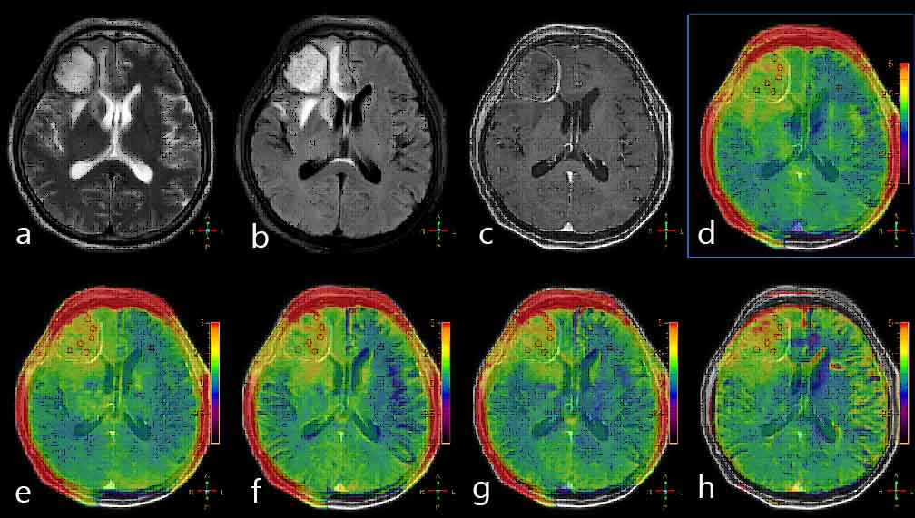
Figure
1a 54-year-old
female patient with meningiomas in brain: a. T2WI; b.
T2WI FLAIR; c. T1-Gd; d. APTw image by SENSE-1.6 fused with T2WI FLAIR image; e-h.
APTw images by CS-SENSE (with factors of 2, 3, 4, and 5) fused with T1-Gd image.
The ROIs were
obtained manually on the SENSE APTw image and copied to the others as shown.
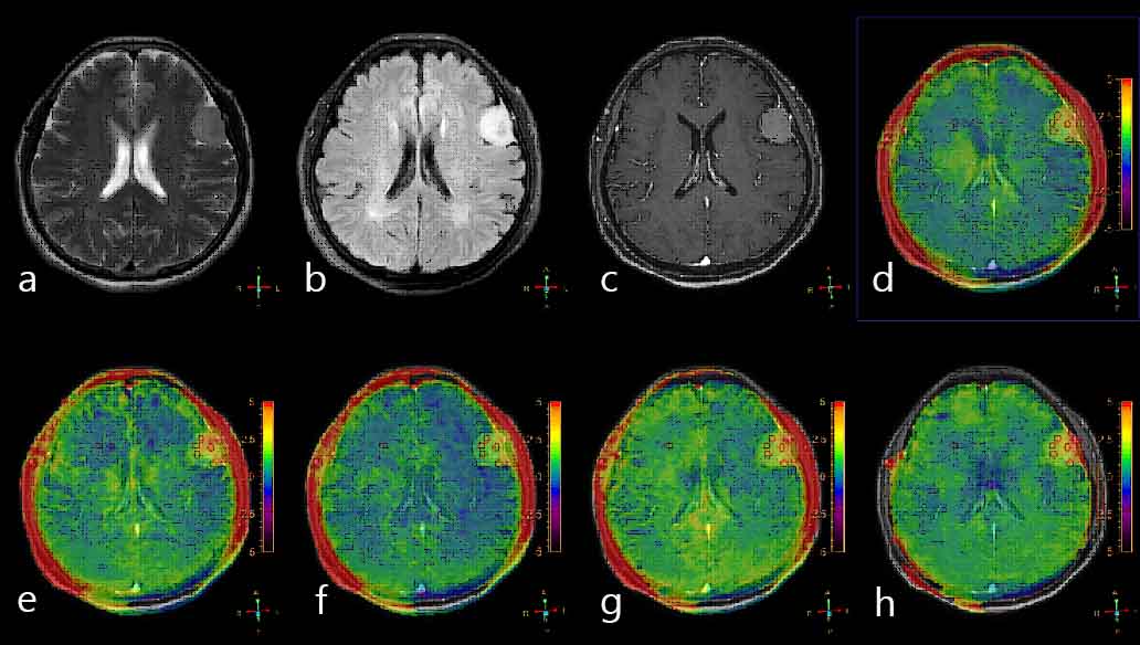
Figure 2 a 64-year-old
female patient with metastatic tumors in brain: a.
T2WI; b. T2WI FLAIR; c. T1-Gd; d. APTw image by SENSE-1.6 fused with T2WI
FLAIR image; e-h. APTw images by CS-SENSE (with factors of 2, 3, 4, and 5) fused
with T1-Gd image. The
ROIs were obtained manually on the SENSE APTw image and copied to the others as
shown