Christoph Stefan Aigner1, Sebastian Dietrich1, Tobias Schaeffter1,2, and Sebastian Schmitter1,3
1Physikalisch-Technische Bundesanstalt (PTB), Braunschweig and Berlin, Germany, 2Division of Imaging Sciences and Biomedical Engineering, King's College London, London, United Kingdom, 3Medical Physics in Radiology, German Cancer Research Center (DKFZ), Heidelberg, Germany
1Physikalisch-Technische Bundesanstalt (PTB), Braunschweig and Berlin, Germany, 2Division of Imaging Sciences and Biomedical Engineering, King's College London, London, United Kingdom, 3Medical Physics in Radiology, German Cancer Research Center (DKFZ), Heidelberg, Germany
This study demonstrates in vivo that 4kT-UP are highly
suitable for calibration-free 3D heart FA homogenization at 7T despite large
inter-subject variations due to varying age, BMI and coil placement.
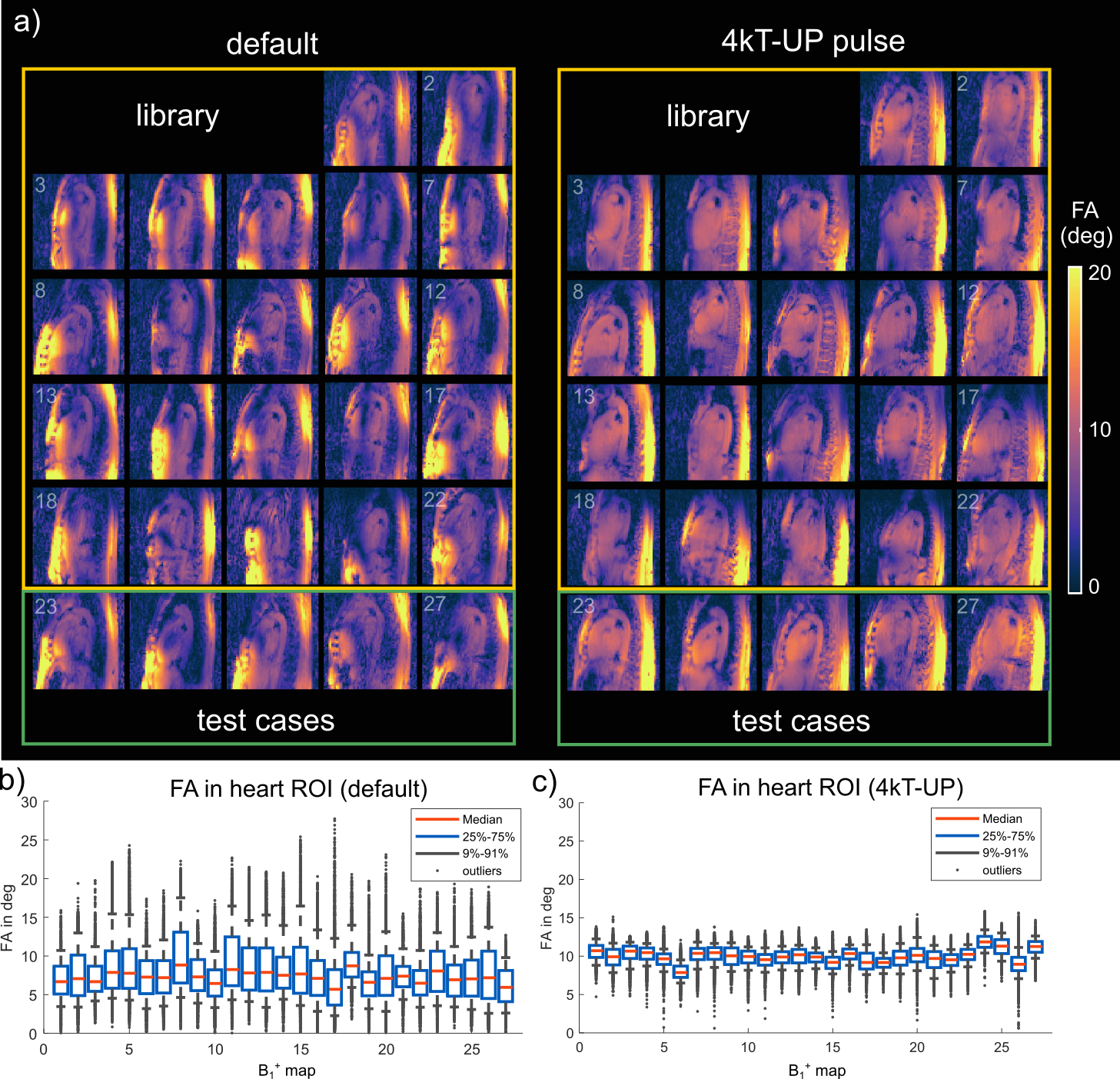
Figure 4: a) Sagittal
slice of the 27 3D B1+ predictions using the default shim setting
(left) and the 4kT-UP (right). Library: B1+ maps 1-22, test-cases: B1+ maps 23-27. The 4kT-UP results in a homogeneous FA in the heart ROI
of all 5 test cases. Moreover, the 4kT-UP achieves a homogeneous FA also in
surrounding tissues such as the aorta. b-c) Boxplot of the FA spread in the 3D
heart ROI of all 27 subjects for default (b) and 4kT-UP (c) demonstrating the
FA homogeneity across all B1+ predictions using the 4kT-UP pulse.
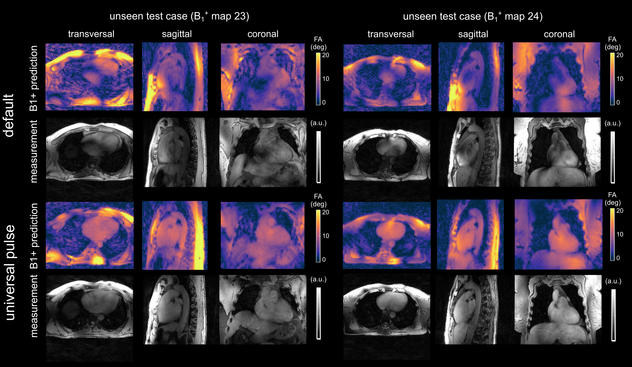
Figure 5: B1+
predictions and reconstructed, respiration corrected 3D GRE images for two unseen test cases with 4kT-UP. These two volunteers were not part of the optimization.
The 3D images are free of breathing artefacts and demonstrate, despite some
differences close to the coil elements, the feasibility to achieve a
homogeneous calibration-free FA of the whole heart. The remaining signal
changes in the AP direction of the acquired images are a result of receive
(B1-) variations.