Thomas Jochmann1, Dejan Jakimovski2, Nora Küchler1, Robert Zivadinov2,3, Jens Haueisen1, and Ferdinand Schweser2,3
1Department of Computer Science and Automation, Technische Universität Ilmenau, Ilmenau, Germany, 2Buffalo Neuroimaging Analysis Center, Department of Neurology at the Jacobs School of Medicine and Biomedical Sciences, University at Buffalo, The State University of New York, Buffalo, NY, United States, 3Center for Biomedical Imaging, Clinical and Translational Science Institute, University at Buffalo, The State University of New York, Buffalo, NY, United States
1Department of Computer Science and Automation, Technische Universität Ilmenau, Ilmenau, Germany, 2Buffalo Neuroimaging Analysis Center, Department of Neurology at the Jacobs School of Medicine and Biomedical Sciences, University at Buffalo, The State University of New York, Buffalo, NY, United States, 3Center for Biomedical Imaging, Clinical and Translational Science Institute, University at Buffalo, The State University of New York, Buffalo, NY, United States
Findings agreed with the existing evidence of iron-caused contrast in deep GM. Findings of larger non-susceptibility frequency in cortical GM compared to WM might stimulate new theories about the biophysical mechanisms of phase contrast in cortical GM.
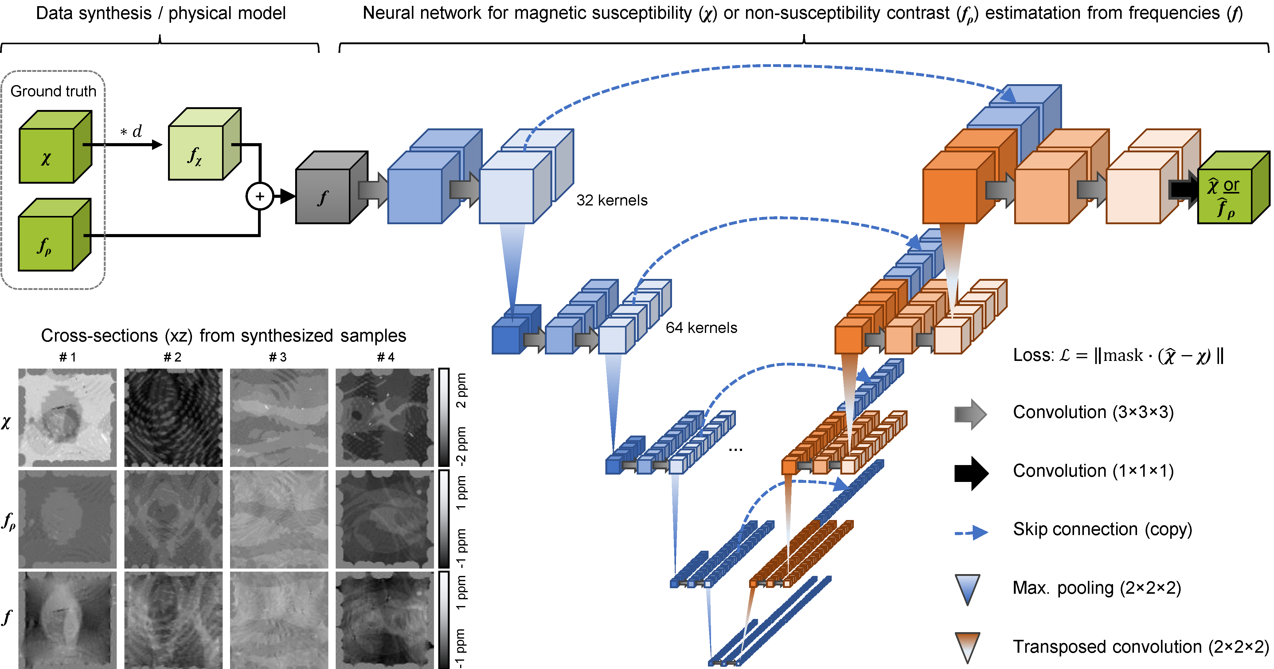
Fig.
1. Physical model for the training data
synthesis, examples of the training data, and the neural network architecture
for mapping the total frequency contrast f
to either of the two source distributions χ or fρ.
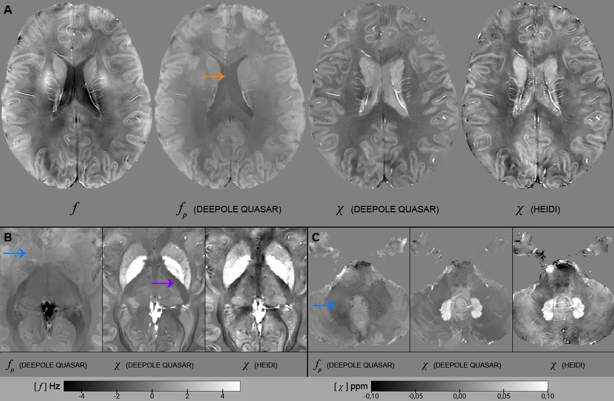
Fig.
4. Three axial slices from a volunteer (f,
33y.). A) fρ has a
stronger positive non-susceptibility frequency shift in gray matter than in
white matter, and cerebrospinal fluid (CSF) has the smallest shift. CSF was
reconstructed homogenously (orange arrow). The iron-rich basal ganglia (B) and
dentate nuclei (C) were isointense in fρ
(blue arrows). Compared to the HEIDI solution, DEEPOLE QUASAR’s χ of the
inner capsule was more similar to surrounding white matter (purple arrow).