Christof Boehm1, Nico Sollmann2,3,4, Jakob Meineke5, Sophia Kronthaler1, Stefan Ruschke1, Michael Dieckmeyer2,3, Kilian Weiss6, Claus Zimmer2,3, Marcus R. Makowski1, Thomas Baum2,3, and Dimitrios C. Karampinos1
1Department of Diagnostic and Interventional Radiology, School of Medicine, Klinikum rechts der Isar, Technical University of Munich, Munich, Germany, 2Department of Diagnostic and Interventional Neuroradiology, School of Medicine, Klinikum rechts der Isar, Technical University of Munich, Munich, Germany, 3TUM-Neuroimaging Center, Klinikum rechts der Isar, Technical University of Munich, Munich, Germany, 4Department of Diagnostic and Interventional Radiology, University Hospital Ulm, Ulm, Germany, 5Philips Research Lab, Hamburg, Germany, 6Philips Healthcare, Hamburg, Germany
1Department of Diagnostic and Interventional Radiology, School of Medicine, Klinikum rechts der Isar, Technical University of Munich, Munich, Germany, 2Department of Diagnostic and Interventional Neuroradiology, School of Medicine, Klinikum rechts der Isar, Technical University of Munich, Munich, Germany, 3TUM-Neuroimaging Center, Klinikum rechts der Isar, Technical University of Munich, Munich, Germany, 4Department of Diagnostic and Interventional Radiology, University Hospital Ulm, Ulm, Germany, 5Philips Research Lab, Hamburg, Germany, 6Philips Healthcare, Hamburg, Germany
The presently proposed water–fat total field inversion method for QSM in water–fat regions showed the following significant improvements over former proposed QSM methods: (a) it significantly reduces background field artifacts, (b) noise amplification and (c) streaking artifacts.
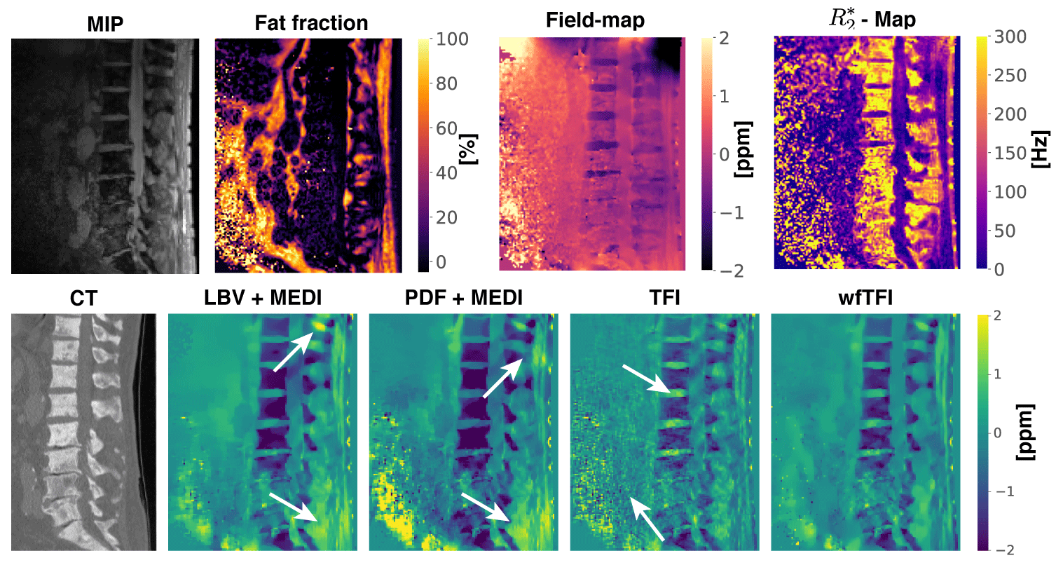
Figure 3: Water–fat imaging and QSM results in a subject with mainly osteoblastic bone metastasis. All 4 QSM methods agree with CT. However, the BFR+LFI methods show BFR artifacts especially in the spinal process region (top arrow) and the fat region in the height of the L5 vertebra (bottom arrow). The TFI method shows significant noise amplification in the anterior to the spinal chord (bottom arrow) and artifactual paramagnetic susceptibility values in all intervertebral discs. The wfTFI shows no noise amplification, no BFR artifacts and no artificial paramagnetic values in the IVDs.
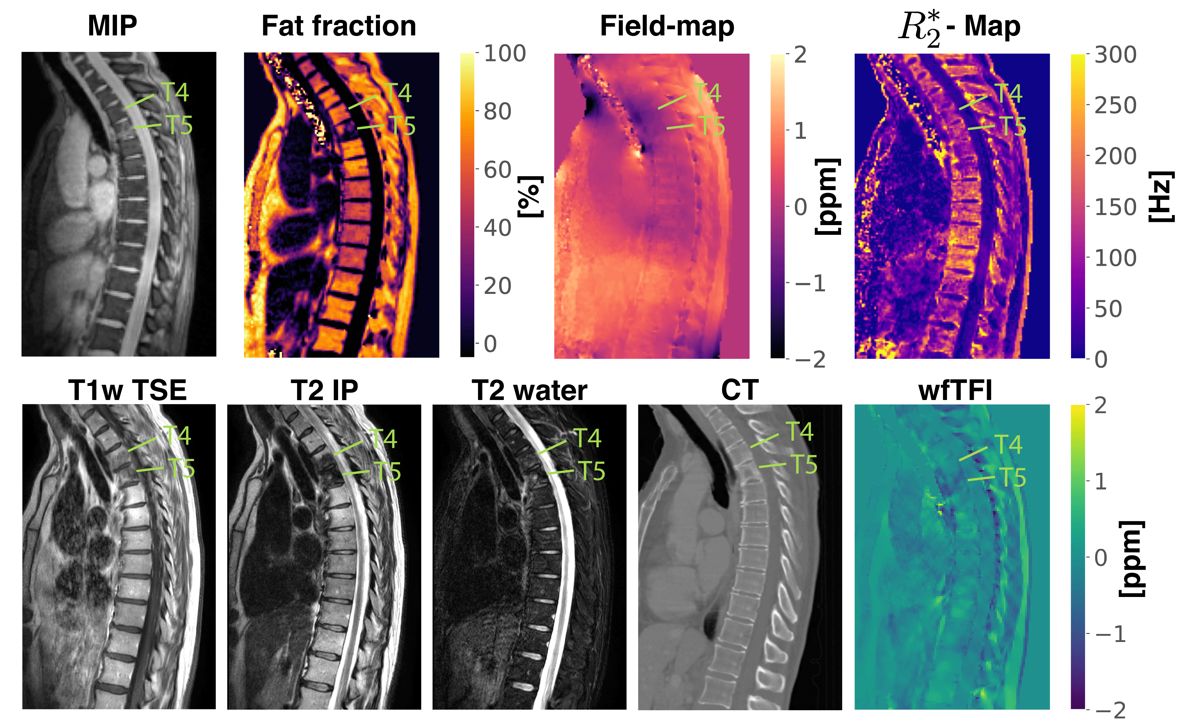
Figure 4: Water–fat imaging (first row), clinical T1w TSE, T2 IP, T2 water, CT and QSM (second row) results in a female patient diagnosed with kidney cancer and mainly osteolytic bone metastases of the vertebrae T4 and T5 according to CT. Yet, the metastatic lesion appears T1- and T2-hypointense, thus suggesting an osteoblastic mass. The results of the wfTFI QSM method, however, suggest an osteolytic pattern, thus being in agreement with CT as the reference standard.