Oliver C. Kiersnowski1, Anita Karsa1, John S. Thornton2, and Karin Shmueli1
1Department of Medical Physics and Biomedical Engineering, University College London, London, United Kingdom, 2UCL Queens Square Institute of Neurology, London, United Kingdom
1Department of Medical Physics and Biomedical Engineering, University College London, London, United Kingdom, 2UCL Queens Square Institute of Neurology, London, United Kingdom
Oblique acquisition
leads to substantial susceptibility errors unless accounted for in QSM susceptibility
calculation. Rotating images to align with B0 and/or defining the dipole kernel along the B0 direction gives accurate susceptibility values.
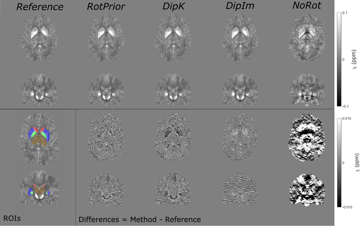
Figure 3: $$$\chi$$$ maps and difference images illustrating the effects of all tilt correction schemes in the numerical phantom. An axial and a coronal slice are shown for a volume tilted at 25° and a reference 0° volume with all $$$\chi$$$ maps calculated using the iterative Tikhonov method. The ROIs analysed are also shown (bottom left). Qualitatively, RotPrior performs the best while NoRot results in substantial $$$\chi$$$ errors across the whole brain. The results from TKD and weighted linear TV (not shown) are very similar.
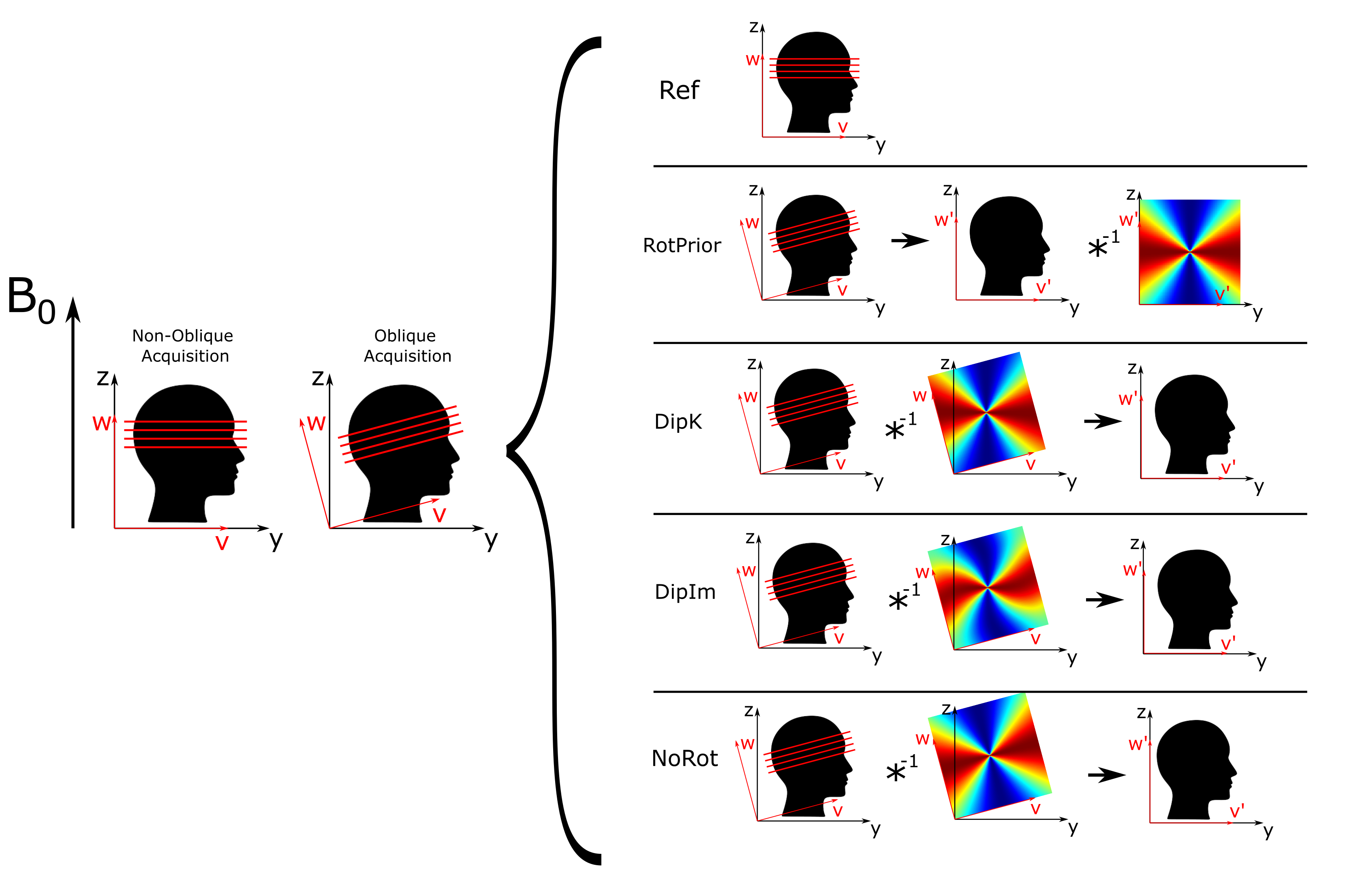
Figure 1: Schematic illustration of oblique acquisition and proposed tilt
correction methods for QSM. The scanner frame (x,y,z) and the
image frame (u,v,w) are shown with respect to the main magnetic
field B0=B0z (left). Proposed tilt correction methods are
shown with the k-space dipole (right). RotPrior involves rotation of the tilted
image frame into
alignment (u',v',w') with the scanner frame. NoRot represents
incorrectly misaligning the dipole kernel with B0z simulating a common
error.