Beata Bachrata1,2, Korbinian Eckstein1, Siegfried Trattnig1,2, and Simon Daniel Robinson1,3,4
1High Field MR Centre, Department of Biomedical Imaging and Image-Guided Therapy, Medical University of Vienna, Vienna, Austria, 2Christian Doppler Laboratory for Clinical Molecular MR Imaging, Vienna, Austria, 3Centre of Advanced Imaging, University of Queensland, Brisbane, Australia, 4Department of Neurology, Medical University of Graz, Graz, Austria
1High Field MR Centre, Department of Biomedical Imaging and Image-Guided Therapy, Medical University of Vienna, Vienna, Austria, 2Christian Doppler Laboratory for Clinical Molecular MR Imaging, Vienna, Austria, 3Centre of Advanced Imaging, University of Queensland, Brisbane, Australia, 4Department of Neurology, Medical University of Graz, Graz, Austria
We address the challenges posed by fat-water chemical shift and relaxation differences in QSM of the head-and-neck at 7T. Using simultaneous fat-water imaging with SMURF and multi-echo information we generate high CNR susceptibility maps free of chemical shift and relaxation effects.

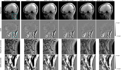
Figure 5: Illustration of the effect of individual chemical shift and relaxation corrections. The susceptibility maps corrected for Type 1 and Type 2 CSA (column 4) clearly depict the paramagnetic fatty areas. The susceptibility maps generated from the conventional images (column 1) and from the SMURF images recombined without corrections for chemical shift effects (column 2 and 3) are more blurred and the values are visibly overestimated. Corrections for T1 and T2* relaxation differences (columns 5 and 6) remove the dominant influence of fat signal in mixed voxels.

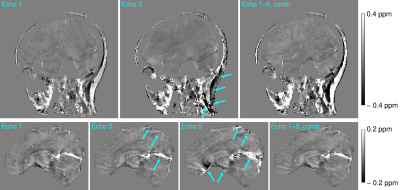
Figure 2: Comparison of QSM maps generated from individual echo images and echo-combined images for the head-and-neck (top) and brain-only (bottom). While in the neck, the first-echo QSM map correctly depicts the paramagnetic fatty areas and provides good CNR (top left), in the brain – with long T2* – the CNR is low. This is improved for later echoes at the expense of short T2* areas, such as the neck, sinuses and veins, where errors occur (centre). In the QSM maps generated from the echo-combined images (right) these errors are reduced and high CNR is achieved over the entire head-and-neck.
