Deepa Darshini Gunashekar1, Lars Bielak1,2, Arnie Berlin3, Leonard Hägele1, Benedict Oerther4, Matthias Benndorf4, Anca Grosu2,4, Constantinos Zamboglou2,4, and Michael Bock1,2
1Dept.of Radiology, Medical Physics, Medical Center University of Freiburg, Faculty of Medicine, University of Freiburg, Freiburg, Germany, 2German Cancer Consortium (DKTK), Partner Site Freiburg, Freiburg, Germany, 3The MathWorks, Inc., Novi, MI, United States, 4Dept.of Radiology, Medical Center University of Freiburg, Faculty of Medicine, University of Freiburg, Freiburg, Germany
1Dept.of Radiology, Medical Physics, Medical Center University of Freiburg, Faculty of Medicine, University of Freiburg, Freiburg, Germany, 2German Cancer Consortium (DKTK), Partner Site Freiburg, Freiburg, Germany, 3The MathWorks, Inc., Novi, MI, United States, 4Dept.of Radiology, Medical Center University of Freiburg, Faculty of Medicine, University of Freiburg, Freiburg, Germany
An explainable deep learning model was implemented to interpret the predictions of a convolution neural network (CNN) for prostate tumor segmentation. The model achieves better visual performance and
fairness for interpreting the decision-making process of the CNN.
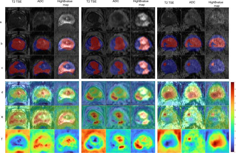
Segmentation of PG and GTV for test patient 1 - 3 (a) Input mpMRI sequences, (b) ground truth, (c) prediction. Overlays in (b) and (c) depict PG in blue and GTV in red.(d-e) overlay of the saliency map for the class PG and GTV on the corresponding input sequences and (f) the generated saliency map PG and GTV. The locations of higher intensity values in the saliency map indicate the importance of the corresponding voxels in the prediction of CNN.
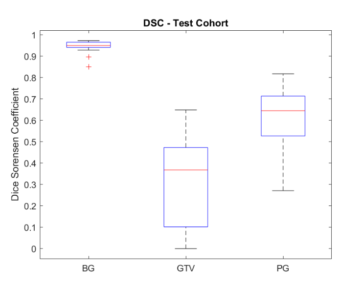
DSC for Test cohort (n = 15). The red lines in the plot show the median Dice value for the classes BG, GTV and PG. The upper and lower bounds of the blue box indicate the 25th and 75th percentiles, respectively.