Chen Cao1,2, Jing Lei2, Yan Gong3, Song Jin2, Jinxia Zhu4, Ming Wei5, and Shuang Xia6
1Department of Radiology, First Central Clinical College, Tianjin Medical University, Tianjin, China, 2Department of Radiology, Tianjin Huanhu Hospital, Tianjin, China, 3Department of Radiology, Tianjin Medical University Nankai Hospital, Tianjin, China, 4MR Collaboration, Siemens Healthcare Ltd., Beijing, China, 5Department of Neurosurgery, Tianjin Huanhu Hospital, Tianjin, China, 6Department of Radiology, Tianjin First Central Hospital, School of Medicine, Nankai University, Tianjin, China
1Department of Radiology, First Central Clinical College, Tianjin Medical University, Tianjin, China, 2Department of Radiology, Tianjin Huanhu Hospital, Tianjin, China, 3Department of Radiology, Tianjin Medical University Nankai Hospital, Tianjin, China, 4MR Collaboration, Siemens Healthcare Ltd., Beijing, China, 5Department of Neurosurgery, Tianjin Huanhu Hospital, Tianjin, China, 6Department of Radiology, Tianjin First Central Hospital, School of Medicine, Nankai University, Tianjin, China
HRMRI determined the cause and length of intracranial large-vessel occlusion, with good agreement with intraoperative angiography. Pre-canalization intraluminal enhancement was higher among patients who received vessel stenting than in those who did not receive stenting.
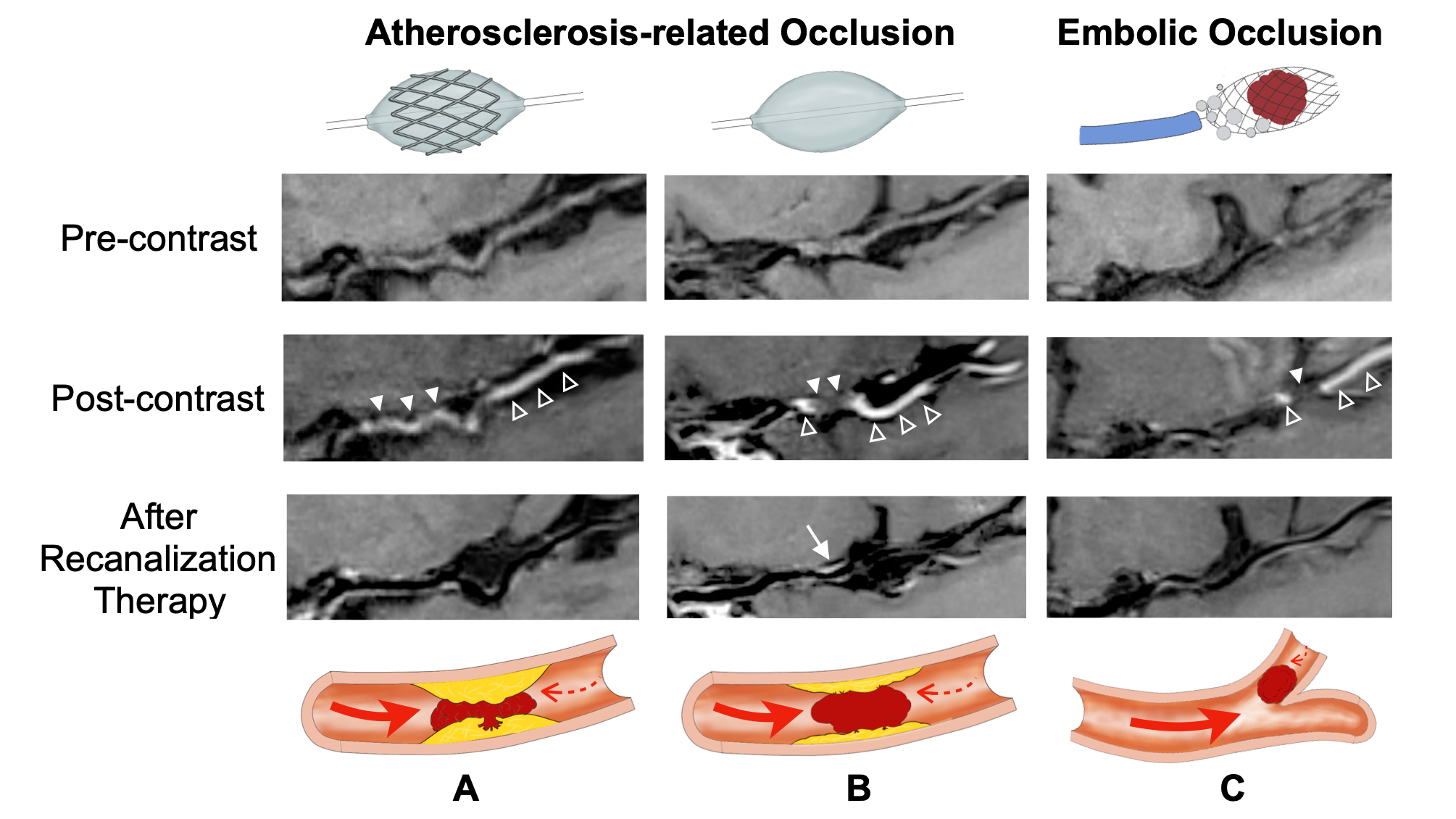
A diagram of acute LVO characteristics and the corresponding typical HRMRI images. HRMRI distinguishes the occluded segments (filled arrowhead) from the slow-flow areas (empty arrowhead). A. ICAS-LVO treated with rescue stenting. Grade 1 enhancement is present on HRMRI, which could reflect higher atherosclerosis plaque percentages compared with those of clots. B. An ICAS-LVO not treated with a stent procedure. Grade 0 enhancement is present on HRMRI, reflecting a higher percentage of clot formation. C. An embolic occlusion treated with a stent retriever.
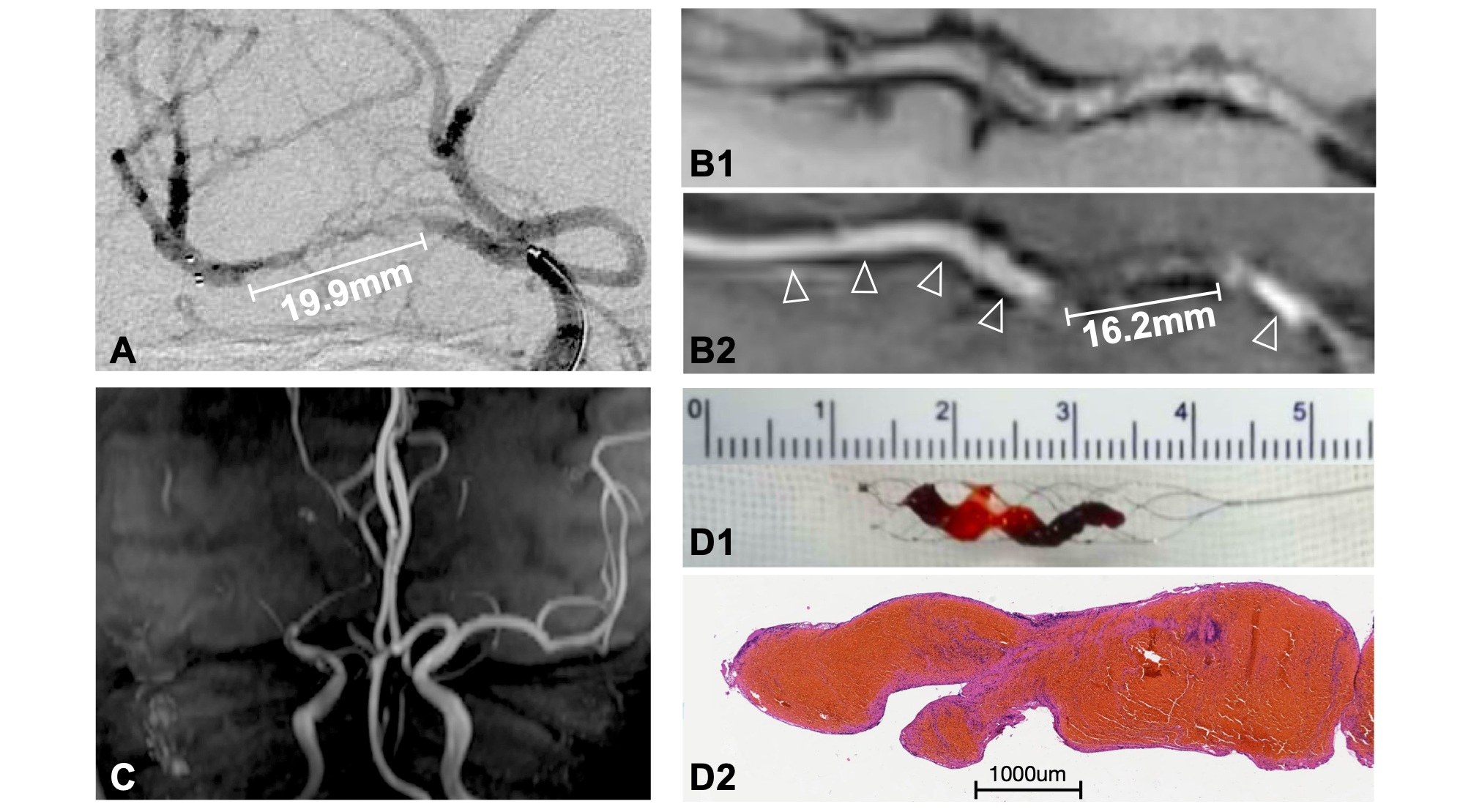
A representative case of occluded segment lengths measured with HRMRI in acute MCA occlusion 7 h after the onset of stroke. The occluded segment lengths measured with angiography (A) and HRMRI (Pre- [B1] and postcontrast [B2] imaging) were 19.9 and 16.2 millimeters, respectively. Slow-flow segments were present at the distal and proximal ends of the occluded segment (arrowhead). (C) The time-of-flight image revealed obstruction of the MCA but did not show the occlusion site specifically. (D1) a gross photograph of a clot. (D2) an H&E-stained section of the clot.