Shannon Helsper1,2, Xuegang Yuan1,2, and Samuel Colles Grant1,2
1National High Magnetic Field Laboratory, Florida State University, Tallahassee, FL, United States, 2Chemical & Biomedical Engineering, FAMU-FSU College of Engineering, Tallahassee, FL, United States
1National High Magnetic Field Laboratory, Florida State University, Tallahassee, FL, United States, 2Chemical & Biomedical Engineering, FAMU-FSU College of Engineering, Tallahassee, FL, United States
23Na
chemical shift imaging and relaxation-enhanced MR spectroscopy at 21.1 T
provides insight into mechanism of ionic and metabolic homeostasis recovery in
cerebral ischemia following hMSC-derived treatments. Methods of EV labeling suitable for in vivo
application are demonstrated.

Fig4. Representative
schematic of Na+/K+ ATPase displays the biological
explanation for utilizing 23Na MRI to assess cerebral ischemia onset
and recovery with treatment. Fractional changes in lesion volume and signal via
23Na CSI provide a sensitive metric to assess treatments.
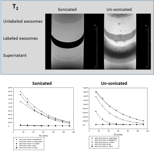
Fig1.
T2 relaxation images and corresponding rate decays
demonstrate SPIO uptake in hMSC EV via sonication and compared to
EV exposed to SPIO but not sonication. Lower layers corresponding to supernatant, with this layer in the un-sonicated sample displaying significant diffusion of SPIO into surrounding gel.