Qingfei Luo1, Zheng Zhong1,2, Kaibao Sun1, and Xiaohong Joe Zhou1,2,3
1Center for MR Research, University of Illinois at Chicago, Chicago, IL, United States, 2Department of Bioengineering, University of Illinois at Chicago, Chicago, IL, United States, 3Departments of Radiology and Neurosurgery, University of Illinois at Chicago, Chicago, IL, United States
1Center for MR Research, University of Illinois at Chicago, Chicago, IL, United States, 2Department of Bioengineering, University of Illinois at Chicago, Chicago, IL, United States, 3Departments of Radiology and Neurosurgery, University of Illinois at Chicago, Chicago, IL, United States
The
scan time of epi-SPEEDI was reduced by ~50% without degrading imaging quality by
using random k-space undersampling and image reconstruction based on joint
spatiotemporal partial separability and sparsity constraints.
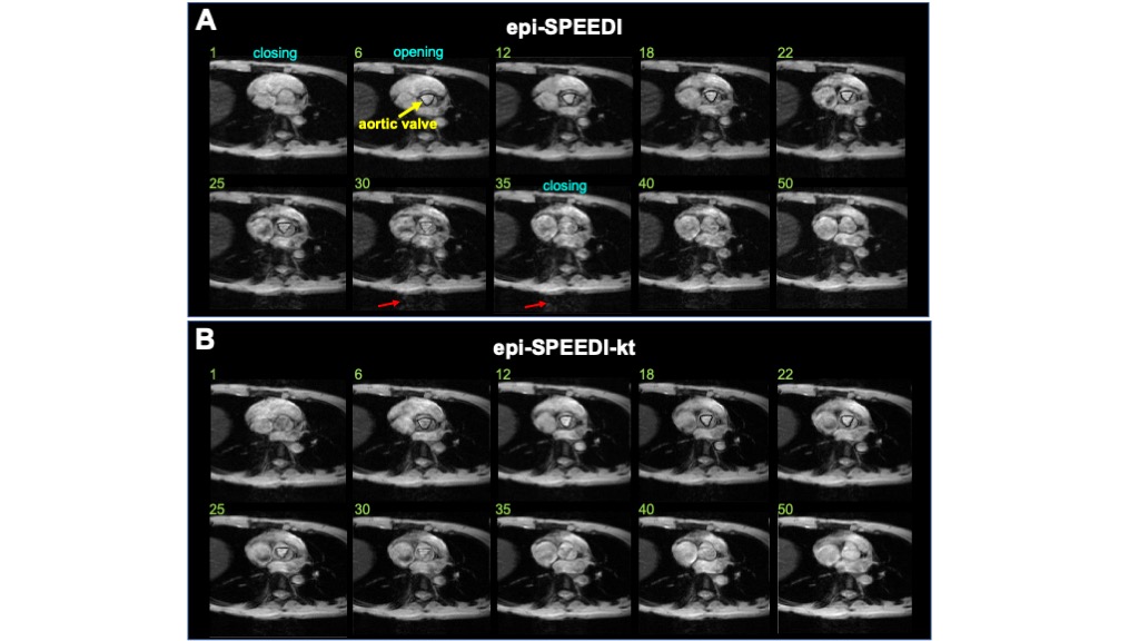
Fig. 4. Dynamic cardiac valve
images acquired using epi-SPEEDI (A) and epi-SPEEDI-kt (B). Each image
corresponds to a specific time point during the aortic valve movement process.
The temporal resolution was 0.6 ms. Images 6-30 show the opening status of
cardiac valve (annotated by the yellow arrow) and the cardiac valve is closed
in the other images. The red arrows indicate artifacts.
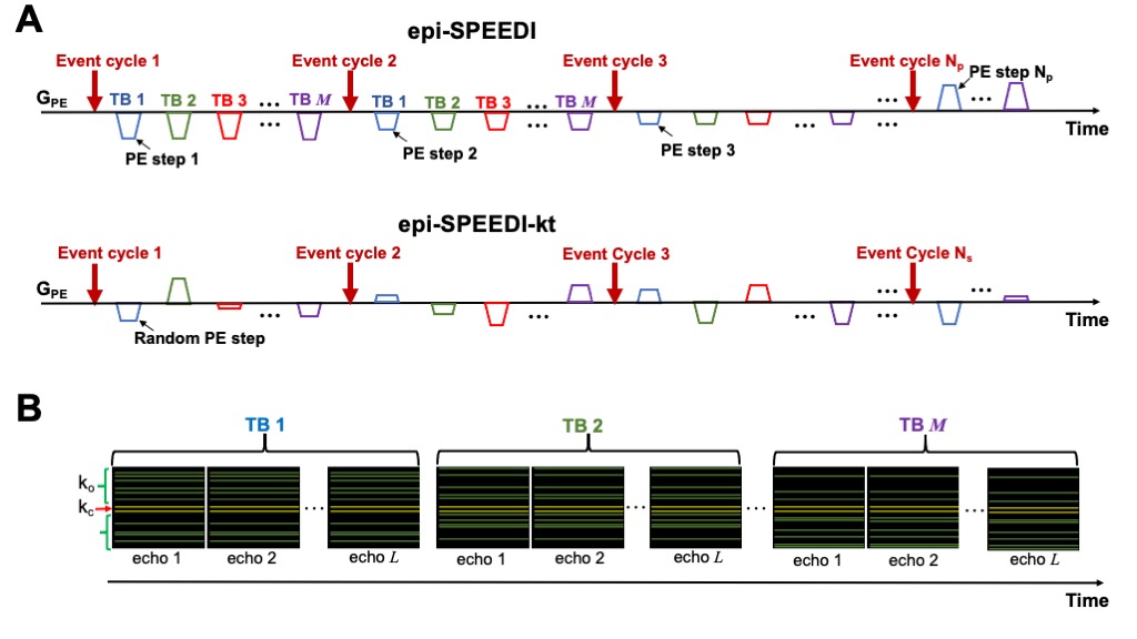
Fig. 2. Comparison of phase-encoding
schemes used in epi-SPEEDI and epi-SPEEDI-kt sequences (A) and the k-space
sampling pattern in epi-SPEEDI-kt (B). Nnav phase lines (yellow
lines) in the central k-space (kc) region are sampled in all the time
blocks (TBs) while the outer k-space (ko) regions are randomly and
sparsely sampled (green lines). The k-space sampling patterns are the same at
the echoes in one TB but different between TBs.